2020 Case Studies – Derm In-Review

December 2020 Case Study

By 2020 Case Studies

December 2020 Case Study

by Kamaria Nelson, MD, MHS

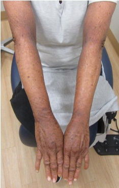
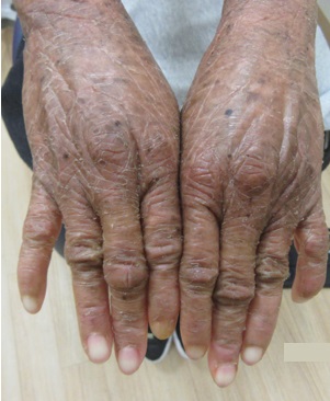
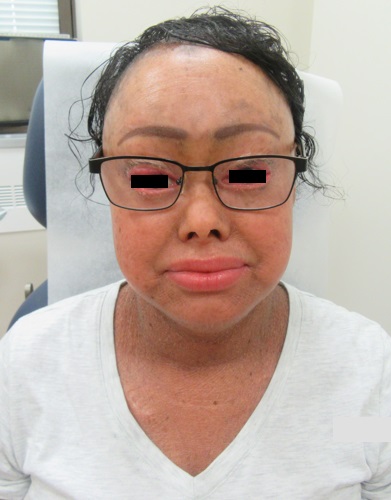
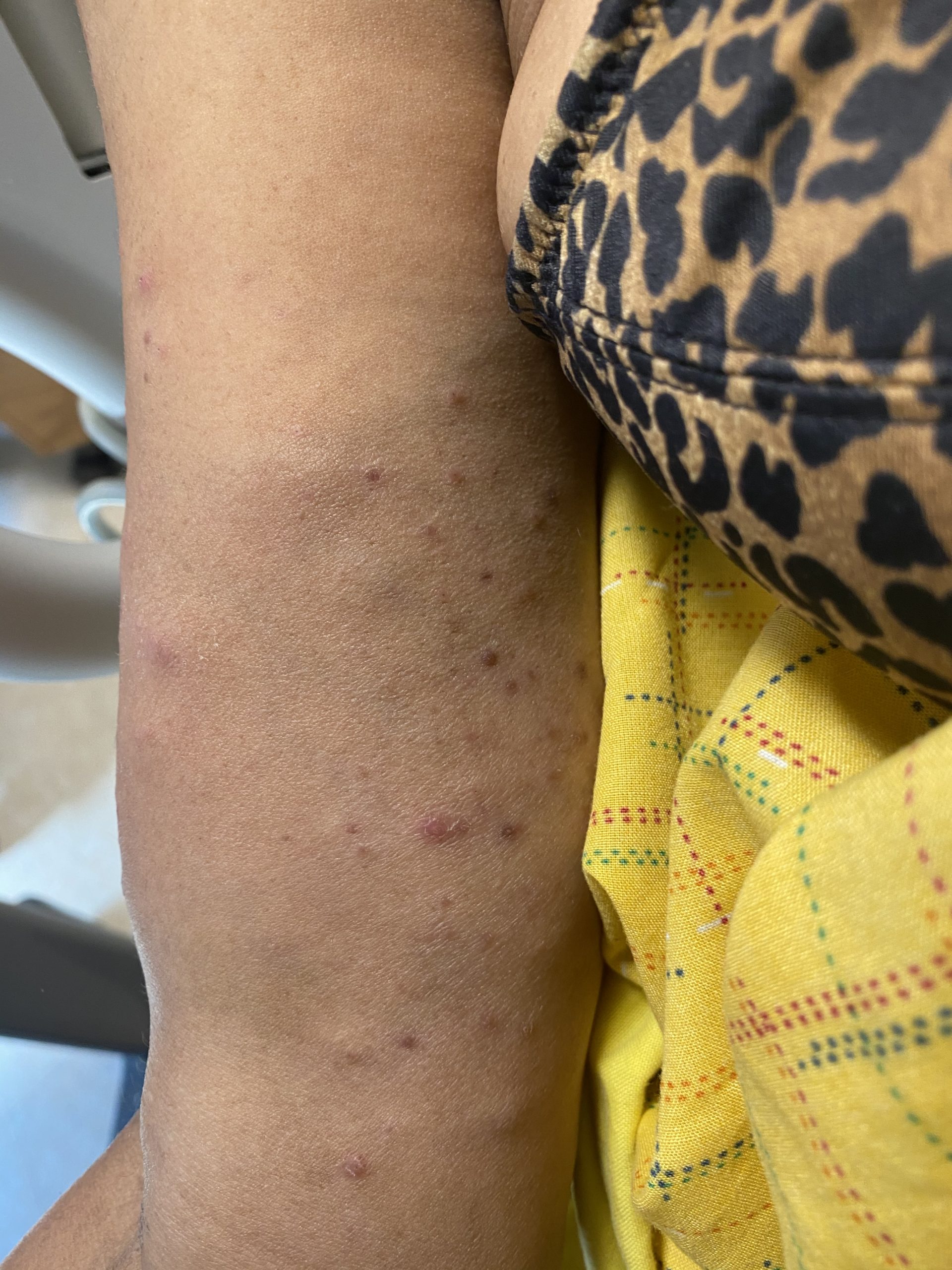
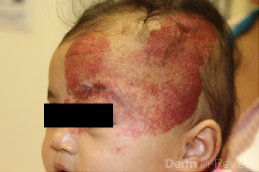
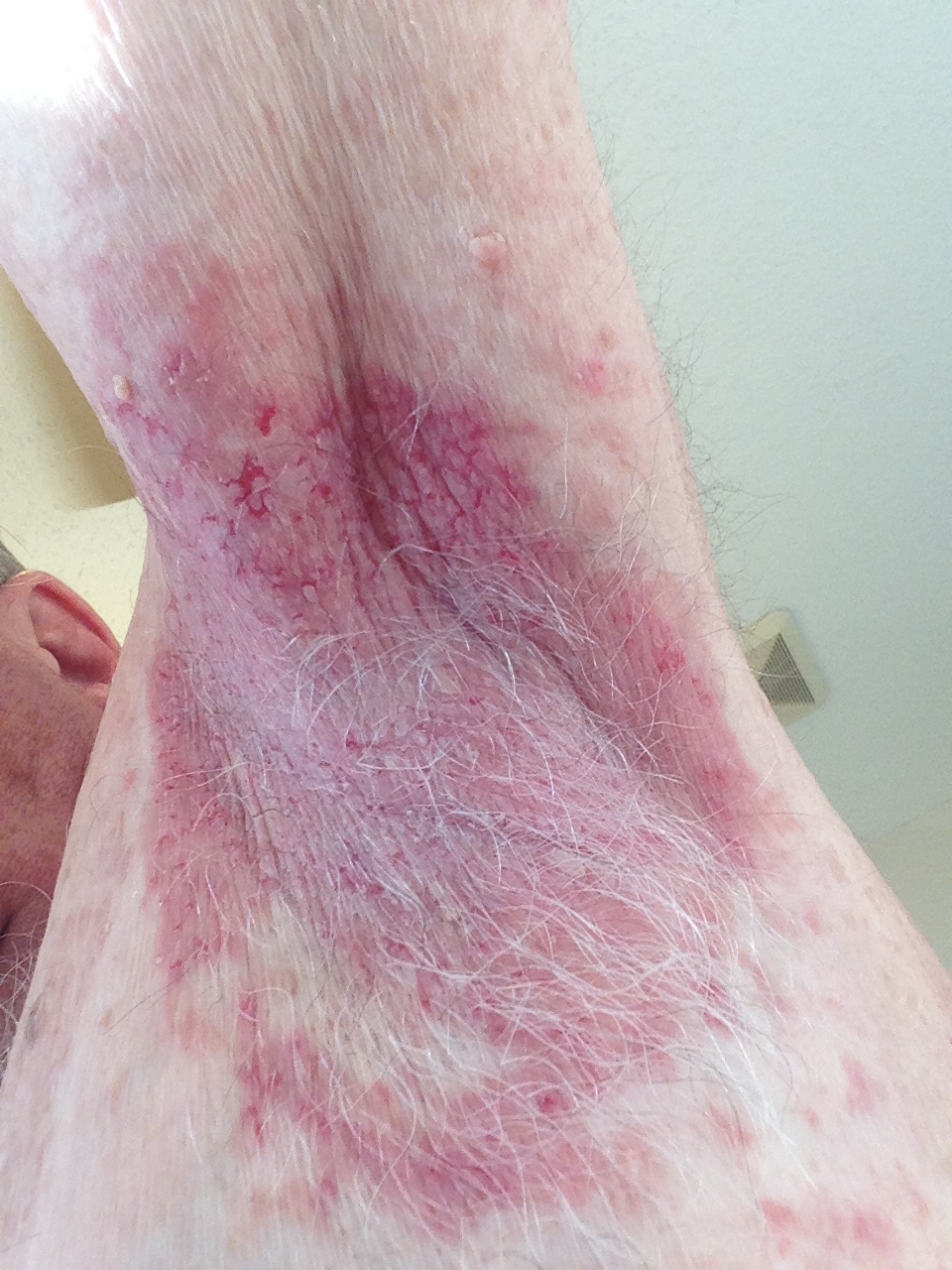
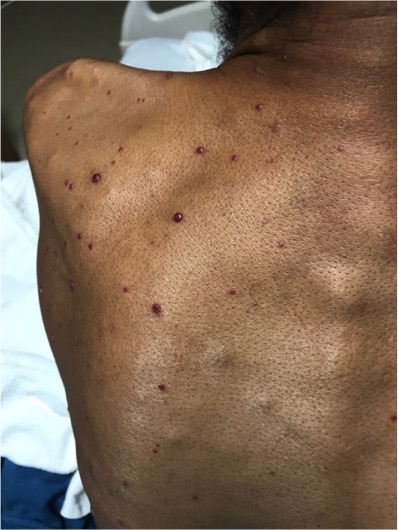
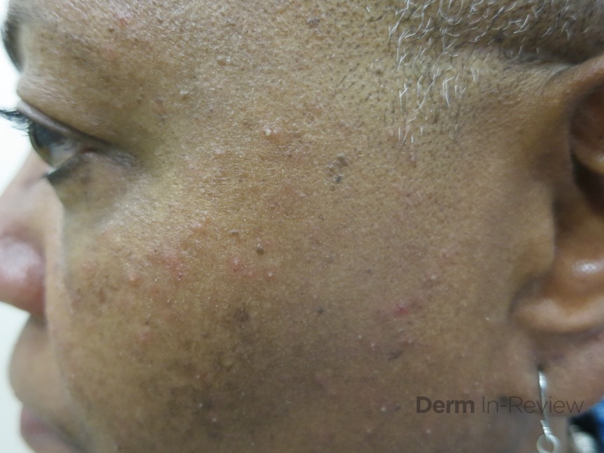
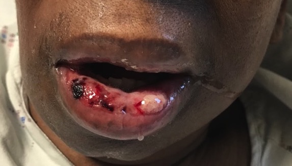
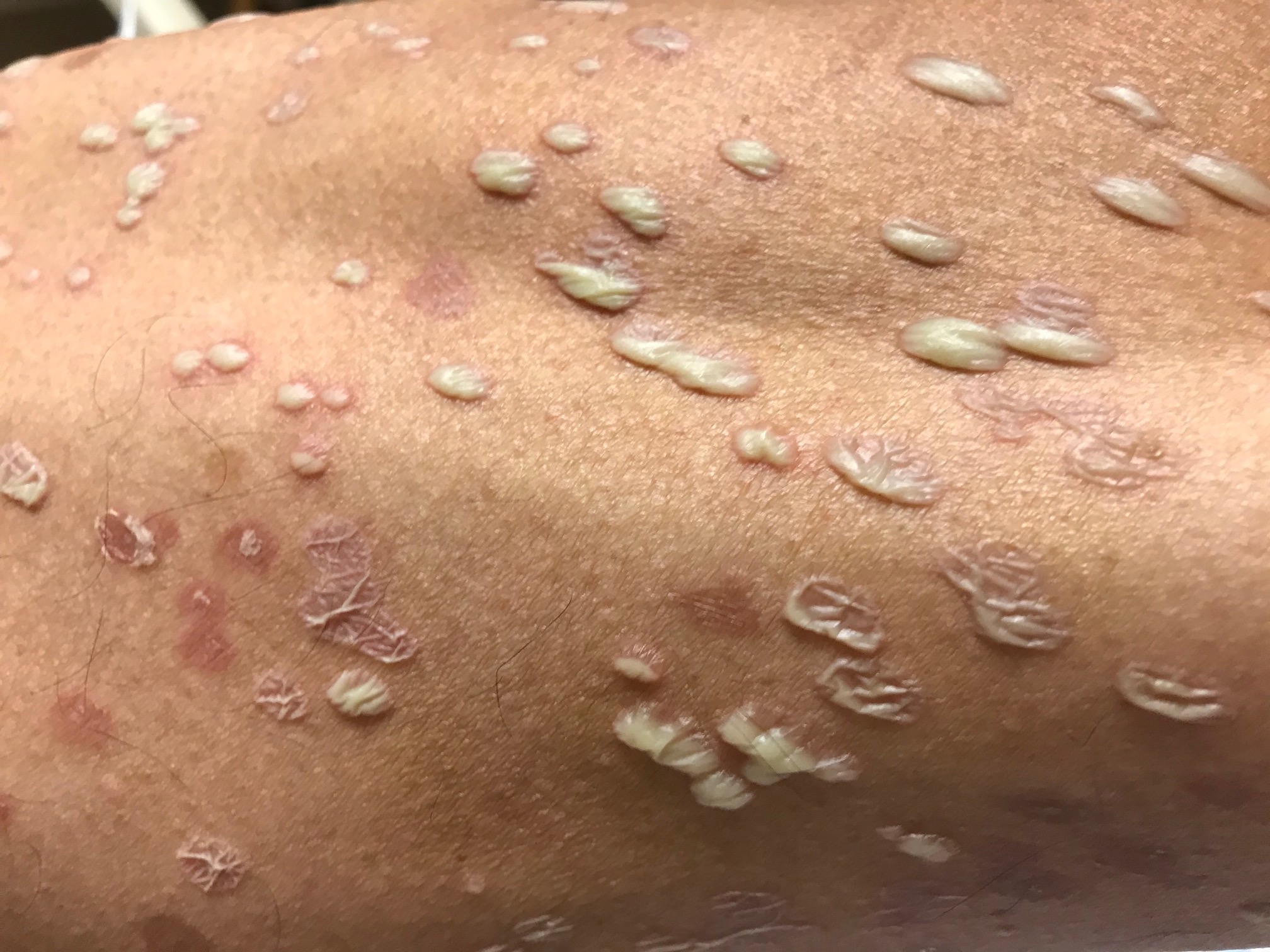
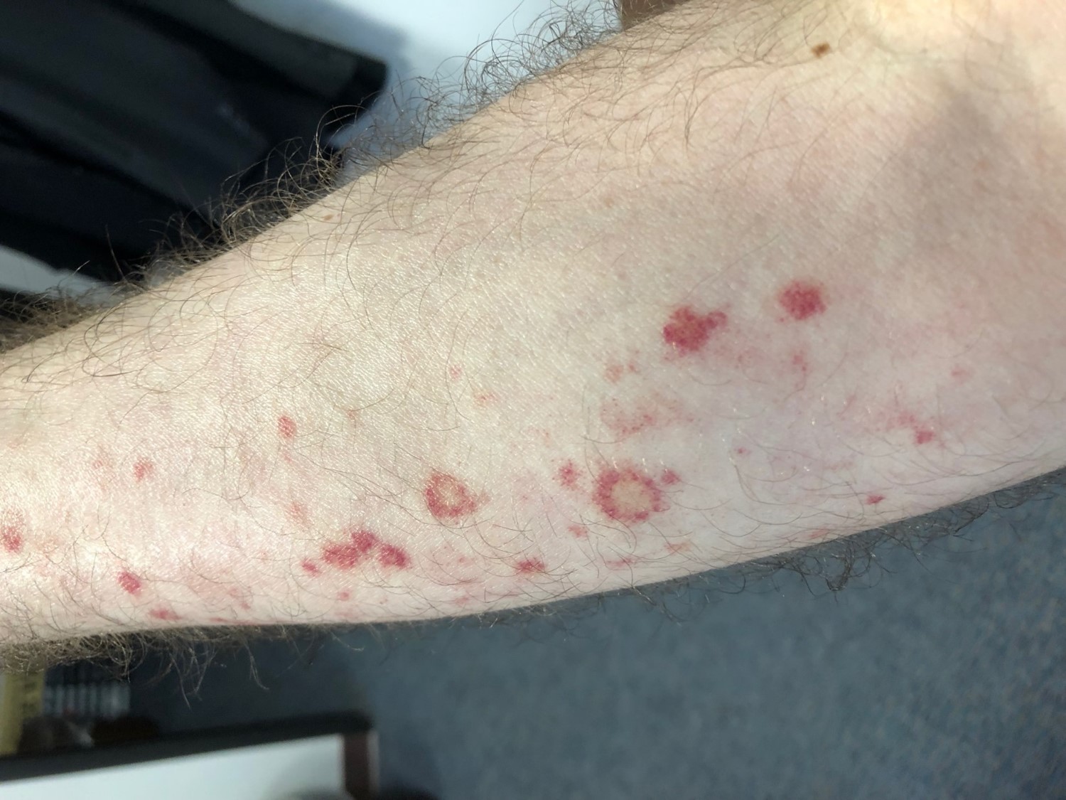
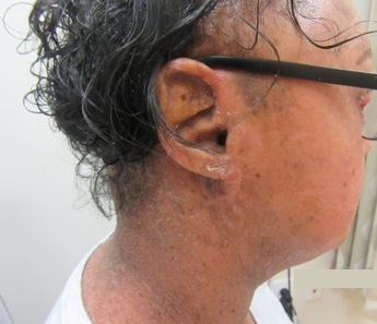
A 33-year-old female presents with a rash that she describes as “thick, excess skin.” The rash has been present since birth. She reports difficulty with regulating her temperature and having dry eyes due to her condition. Current treatments include urea, petroleum jelly, and n-acetyl cysteine. She was on oral acitretin from ages 3-14. The patient reports no other history of cutaneous disease or systemic conditions. She has a sister with similar skin findings. Physical examination reveals thick, plate-like, dark appearing scaly plaques on the arms, upper chest, neck and face; ectropion and dysmorphic appearing facies were present. (Images 1-4).

Based upon the patient history and clinical examination, which genetic mutation is most likely related to this condition?

A.) ALDH3A2 (or ALDH10)
B.) TGM1
C.) Genes encoding KRT1 & KRT10
D.) ABCA12
E.) STS

Correct answer: B) TGMI

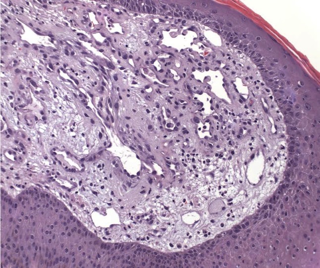
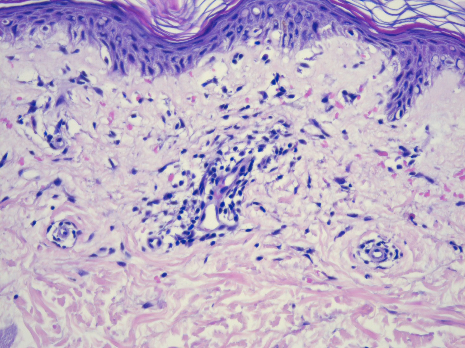
Lamellar ichthyosis (LI) in an autosomal recessive congenital ichthyosis characterized by diffuse large, gray brown scales that are quadrilateral and can resemble an armor plate. During the first 2-3 weeks of life, LI frequently presents with a collodion membrane with ectropion that then desquamates. 35-55% of all patients have transglutaminase-1 deficiency due to mutations in TGM1. Transglutaminase-1 is found in the upper layers of the epidermis where it catalyzes the linking of proteins and aids in the formation of ε-lysine isopeptide bonds and ester bonds between proteins and ω-hydroxyceramides.

Transglutaminase-1 crosslinks structural proteins to each other to form the cornified cell envelope. The formation of the cornified cell envelope is important for intracellular lipid layer formation in the stratum corneum; therefore, mutations can cause defective barrier function. Mutations in CYP4F22 results in an acral, milder presentation of lamellar ichthyosis. CYP4F22 encodes an enzyme that’s responsible for ω-hydroxylation of ultra-long-chain fatty acids which form acyleramides, important to skin barrier function. Histologic features are not diagnostic and may reveal orthokeratotic hyperkeratosis that covers an acanthotic epidermis, normal epidermal proliferation rate, and a thin or absent cornified cell envelope on electron microscopy. Treatment consists of adequate moisturization with emollients. Topical retinoids can be used along with ammonium lactate and urea for adherent scale. For more severe cases, systemic retinoids, such as isotretinoin or acitretin, should be considered.

Sjogren-Larsson syndrome (SLS) is an autosomal recessive neurocutaneous disorder that is caused by mutations in ALDH3A2, which leads to a deficiency of fatty aldehyde dehydrogenase (FALDH). FALDH catalyzes oxidation of long-chain aliphatic aldehydes into fatty acids; mutations lead to accumulation of fatty alcohol/aldehyde-modified lipids and proteins causing membrane alteration. SLS is characterized by congenital ichthyosis, spastic paralysis, intellectual disability, and degenerative retinitis. Patients present with a juvenile macular dystrophy exhibited by perifoveal glistening white dots in the ocular fundus which are present during the first year of life.  SLS is also associated with persistent pruritus, unlike the other ichthyoses.

Epidermolytic ichthyosis (EI), also known as epidermolytic hyperkeratosis, is an autosomal dominant disorder that is caused by heterozygous mutations in the genes that code for keratin 1 (KRT1) and keratin 10 (KRT10). KRT1 and KRT10 are expressed in the differentiated spinous and granular layers of the epidermis; mutations compromise epidermal integrity leading to cytolysis and blistering. EI usually presents with blisters at or shortly after birth and later, thick, uniquely malodorous spine-like scale on the flexures. Key histological findings include orthokeratotic hyperkeratosis, prominent acanthosis, hypergranulosis and cytolysis of suprabasal and granular layers; keratinocytes reveal intracellular vacuolization and dense clumps of keratin intermediate filaments (KIFs).

Harlequin ichthyosis (HI) is rare and the most severe form of autosomal recessive congenital ichthyosis. It is caused by loss-of-function mutations in ABCA12. ABCA12 is responsible for ceramide and lipid transport into lamellar bodies; mutations prevent the formation of lipid bilayers in the stratum corneum resulting in hyperkeratosis and a severe breakdown of the barrier. HI presents with thick collodion, diffuse armor-like plates and severe ectropion and eclabium. Patients with HI are usually born premature and have a high mortality rate (nearing 50%) due to respiratory insufficiency from severe immobilization caused by the thickened stratum corneum or sepsis from severely fissured skin.

X-linked ichthyosis results from a deficiency of steroid sulfatase caused by the deletion of the STS gene on chromosome Xp22.31. This results in inadequate hydrolysis of cholesterol sulfate and dehydroepiandrosterone sulfate (DHEAS) causing accumulation of cholesterol 3-sulfate in the epidermis which can inhibit tranglutaminase-1. Women who are pregnant with an affected fetus often have failure to initiate labor due to inadequate conjugation of DHEAS, necessary for estrogen synthesis. At birth, males may present with erythroderma and exfoliation of large, translucent scale. This later progresses to polygonal, dark-brown plates on the neck, extremities and trunk; periauricular involvement is characteristic.  Other important findings include asymptomatic corneal opacities, increased incidence of cryptorchidism and risk for testicular cancer, and higher prevalence of attention deficit disorder. Molecular analysis (genome microarrays, FISH, PCR) can detect the genetic defect and may be used prenatally from amniotic samples. Noninvasive techniques include serum estriol levels and urine non-hydrolyzed sulfated steroids or biochemical assay to measure steroid sulfatase enzyme activity. Plasma cholesterol sulfate levels after birth can be used to aid in diagnosis.

References

  1. Bolognia, J., Schaffer, J., & Cerroni, L. (2019). Dermatology (Fourth edition.). Elsevier.
  2. James, W., Elston, D., Treat, J., Rosenbach, M., Neuhaus, I., & Andrews, G. (2013). Andrews’ Diseases of the Skin : Clinical Dermatology (Thirteenth edition.). Elsevier.

November 2020 Case Study

By 2020 Case Studies

November 2020 Case Study

by Azam Qureshi, MD

A 62-year-old female presents with a three week history of rash affecting the upper and lower extremities. On physical examination, subcutaneous nodules and multiple pink to purple scattered papules are noted on the distal upper extremities and the lower extremities extending from the outer thighs to the mid-anterior thighs bilaterally (Figures 1 & 2). The patient reports that some of the subcutaneous nodules are tender. She has not used any previous treatments. She has no family history of chronic diseases, including skin or autoimmune disease. She reports recent shortness of breath and mild cough, but otherwise, has been feeling well with no fevers, chills, night sweats, unexplained weight loss, abdominal pain, nausea, vomiting, diarrhea, headaches, or joint pains.

Blood work was notable for elevated erythrocyte sedimentation and chest x-ray was notable for only bilateral hilar lymphadenopathy. Two separate four millimeter punch biopsies were performed, and showed non-caseating epithelial granulomas throughout the superficial and deep dermis which lacked significant inflammatory rim of lymphocytes or plasma cells. Star-shaped eosinophilic inclusions of collagen were also seen. Brown-Hopps, Fite, and Gomori methenamine silver special stains were conducted on both biopsies and were negative for microorganisms.

Further investigation into the patient’s medical history is most likely to provide evidence for which of the following?

A.) Recent treatment of Hepatitis C with sofosbuvir & ledipasvir
B.) Recent spelunking in Kentucky
C.) Recent initiation of anti-retroviral therapy for treatment for HIV
D.) Recent travel from India
E.) History of major bowel surgery

Correct answer: C) Recent initiation of anti-retroviral therapy for treatment for HIV

This patient is presenting with sarcoidosis, as evidenced by the cutaneous morphologies of subcutaneous nodules and pink to purple papules affecting the upper and lower extremities, elevated erythrocyte sedimentation rate, pulmonary symptoms with bilateral hilar lymphadenopathy noted on chest x-ray, and skin biopsies showing evidence of “naked” epithelioid granulomas and asteroid bodies with negative Brown-Hopps, Fite, and Gomori methenamine silver special stains.

Anti-retrovirals in HIV patients have been shown to lead to possible drug-induced sarcoidosis.1,2 Furthermore, development of cutaneous sarcoidosis has been described as a feature of immune reconstitution syndrome in HIV patients initiating antiretroviral therapy.2 Although ribavirin and interferon-α treatment for hepatitis C have been associated with sarcoidosis onset, no such association has been made with the hepatitis C treatments of sofosbuvir or ledipasvir.3,4

Histoplasmosis (B) and cutaneous tuberculosis (D) may also present with pulmonary symptoms in conjunction with cutaneous manifestations and skin biopsy findings showing granulomatous inflammation. Sarcoidosis is the more likely diagnosis in this patient, as histoplasmosis presents more commonly with cutaneous molluscoid nodules, cellulitis, ulcers, panniculitis, and oral lesions. Skin biopsy would show intracellular 2-4 micron yeast with a surrounding halo within histiocytes. Cutaneous tuberculosis can present in a diverse manner, with histopathology showing evidence of suppurative inflammation, later becoming granulomatous with central caseous necrosis.

Cutaneous Crohn’s disease (E) can present with manifestations such as erythema nodosum, pyoderma gangrenosum, pyostomatitis vegetans, epidermolysis bullosa acquisita, and acrodermatitis enteropathica. There may be genital, perianal, or oral involvement. Cutaneous Crohn’s occurring away from the perianal, genital, or oral areas is rare, and more often affects the lower extremities and soles of the feet, with less common involvement of the upper extremities. Histopathology often shows non-caseating tuberculoid granulomas with an inflammatory rim of lymphocytes in the superficial and deep dermis, along with frequent Langerhans giant cells.

References

  1. Pascual JC, Belinchon I, Silvestre JF, Vergara G, Blanes M, Bañuls J, Betlloch I, Boix V. Sarcoidosis after highly active antiretroviral therapy in a patient with AIDS. Clinical and Experimental Dermatology: Clinical dermatology. 2004 Mar;29(2):156-8.
  2. Rachadi H, Ramli I, Meknassi I, Hassam B, Benzekri L. Recurrence of cutaneous sarcoidosis during immune reconstitution syndrome in an HIV-infected patient. InAnnales de dermatologie et de venereologie 2015 Dec (Vol. 142, No. 12, pp. 757-760).
  3. Hurst EA, Mauro T. Sarcoidosis associated with pegylated interferon alfa and ribavirin treatment for chronic hepatitis C: a case report and review of the literature. Archives of Dermatology. 2005 Jul 1;141(7):865-8.
  4. Cogrel O, Doutre MS, Marliere V, Beylot‐Barry M, Couzigou P, Beylot C. Cutaneous sarcoidosis during interferon alfa and ribavirin treatment of hepatitis C virus infection: two cases. British Journal of Dermatology. 2002 Feb;146(2):320-4.
  5. Bolognia JL, Schaffer JV, Duncan KO, Ko CJ. Dermatology essentials E-book. Elsevier Health Sciences; 2014 Feb 26.

October 2020 Case Study

By 2020 Case Studies

October 2020 Case Study

by Blair Allais, MD

A 2-month-old female infant, born prematurely with a birth weight less than <1000g, presents to dermatology clinic with the following findings. No family history of similar. Physical examination revealed a large hemangioma > 5cm on the face including the scalp.

Which of the following is true regarding her condition?

A.) GNAQ mutations have been identified in these patients
B.) This patient is at risk for high output cardiac failure
C.) Evaluation with head computed tomography (CT) scan typically reveals cortical “tram track” calcifications
D.) This patient is at increased risk for spinal dysraphism
E.) MRI/MRA of the head and neck is indicated to evaluate for anomalies of major cerebral or cervical arteries

Correct answer: E) This patient has PHACE(S) syndrome, and MRI/MRA of the head and neck is indicated to evaluate for cerebrovascular anomalies.

A) False: Somatic mosaic mutations in the GNAQ gene have been described in Sturge-Weber syndrome, which is characterized by capillary malformations in a V1 and V2 > V3 distribution.

B) False: High output cardiac failure is a clinical finding in diffuse neonatal hemangiomatosis (> 5 hemangiomas with organ involvement), kasabach-merritt phenomenon (association with tufted angiomas or kaposiform hemangioendotheliomas) and klippel-trenaunay syndrome (characterized by capillary malformation, venous malformation, and/or lymphatic malformation of a limb).

C) False: Tram-track calcifications are often described on head CTs of patients with sturge-weber syndrome.

D) False: spinal dysraphism is part of the constellation of findings in LUMBAR/SACRAL syndrome.

E) True: This patient has PHACE(S) syndrome, and MRI/MRA of the head and neck is indicated to evaluate for cerebrovascular anomalies.

This infant has PHACE(S) syndrome, which was first described in 19961 for the following spectrum of findings: P, posterior fossa and other structural brain malformations; H, hemangioma; A, arterial anomalies of cervical and cerebral vessels; C, cardiac defects (especially coarctation of the aorta); E, eye anomalies (retinal vascular anomalies, optic nerve hypoplasia); and S, sternal defects and supraumbulical raphe.2 In a study of 108 infants with large facial hemangiomas (>22cm), 31% had PHACES syndrome and 91% of affected individuals had more than one extracutaneous manifestation. The most common findings were cerebrovascular, cardiovascular, and structural brain abnormalities.3 In patients with segmental cervicofacial hemangiomas, PHACE(S) syndrome must be excluded with MRI/MRA of the head and neck, ophthalmologic exam and echocardiogram/cardiac evaluation.

References

Dermatology In Review, Image Companion Figure 3.8.3 PHACES
1.) Frieden IJ, Reese V, Cohen D. PHACE syndrome: the association of posterior fossa abnormalities, arterial anomalies, coarctation of the aorta and cardiac defects, and eye abnormalities. Ach Dermatol 1996; 132:307-11.
2.) Haggstrom AN, Garzon MC. Infantile Hemangiomas. In: Dermatology , Edited by Jean L. Bolognia , Julie V. Schaffer , Lorenzo Cerroni Fourth edition , China: Elsevier, 2018, ISBN 978–0‐7020–6275–9
3.) Haggstrom AN, Garzon MC, Baselga E et al. Risk for PHACE syndrome in infants with large facial hemangiomas. Pediatrics 2010; 126:e418-26

September 2020 Case Study

By 2020 Case Studies

September 2020 Case Study

by Adrianna Gonzalez, MD

A 57-year-old male was referred to the outpatient dermatology clinic for the evaluation of a 9-year history of recurrent pruritic and painful vesicles and erosions of bilateral axillae. He had been treated in the past by his primary care physician with topical and oral steroids, as well as topical antibiotics with only mild temporary improvement of symptoms and in the appearance of the lesions. Patient had a history of hyperlipidemia but denied history of any other cutaneous or autoimmune conditions. Patient was adopted and had no information on family history. Physical examination revealed macerated erythematous plaques with fissuring and erosions on bilateral axillary vaults.

Based on the history and physical exam, which of the following is the most likely diagnosis?

A.) Darier disease
B.) Hailey-Hailey disease
C.) Candidiasis
D.) Granular parakeratosis
E.) Intertrigo

Correct answer: B. Hailey-Hailey disease

Hailey-Hailey disease (HHD), also known as benign familial pemphigus, is a rare autosomal dominant intraepidermal blistering disorder characterized by flaccid vesicles or bullae in a circinate pattern, arising on intertriginous areas such as the neck, axillae, inframammary region, perineum and inguinal folds. Clinically, patients most often present with painful and pruritic erythematous macerated plaques with fissures and crusting that can frequently be superinfected and malodorous. Onset is typically between the second and fourth decades of life and patients often experience a variable course with relapses and remissions. Relapses tend to recur at sites of prior involvement and lesions tend to be exacerbated by heat, sweat, friction and stress.  Lesions typically heal without scarring but often cause post-inflammatory hyperpigmentation.

HHD is caused by a mutation in the ATP2C1 gene which encodes a calcium-dependent adenosine triphophatase responsible for regulating the sequestration of calcium in the Golgi apparatus of epidermal cells. Defect in this protein leads to impaired function of desmosomes, which causes keratinocyte adhesion defects and acantholysis. Histopathology shows acanthosis and full-thickness acantholysis resembling a “dilapidated brick wall.”

Management of HHD can be very difficult. Loose clothing and prevention of sweating and friction is always recommended for prevention. For mild cases, topical corticosteroids and topical antibiotics may be effective. Oral antibiotics, intralesional corticosteroids or systemic corticosteroids may sometimes be needed. For recalcitrant cases, numerous treatment modalities have been used with varying success such as topical calcineurin inhibitors, methotrexate, cyclosporine, oral retinoids, botulinum toxin, photodynamic therapy (PDT), terbinafine, dapsone, carbon dioxide laser ablation, cryotherapy, dermabrasion, naltrexone and narrow-band ultraviolet B therapy (NBUVB), among others.

Among the other answer choices, Darier disease normally presents earlier in life and lesions typically start as red-brown papules or plaques rather than vesicles or bullae. Darier disease may also have mucosal involvement while HHD does not. Intertrigo can resemble HHD, however these erythematous patches are commonly seen in the intertriginous areas of obese individuals and will lack the family history typically seen in HHD. Candidiasis has a more beefy-red appearance and typically has satellite pustules. Granular parakeratosis most commonly presents as red to brown hyperkeratotic papules that can coalesce into well-demarcated plaques and is typically seen in the axillae of females.

References

  1. Alajmi A, Jfri A, Lovett A. Hailey-Hailey disease treated successfully with naltrexone and magnesium. JAAD Case Rep. 2019;5(9):760-762.
  2. Albers LN, Arbiser JL, Feldman RJ. Treatment of Hailey-Hailey Disease With Low-Dose Naltrexone. JAMA Dermatol. 2017;153(10):1018-1020.
  3. Engin B, Kutlubay Z, Celik U, Serdaroglu S, Tuzun Y. Hailey-Hailey disease: A fold (intertriginous) dermatosis. Clin Dermatol. 2015;33(4):452-455.
  4. Yadav N, Madke B, Kar S, Prasad K, Gangane N. Hailey-Hailey disease. Indian Dermatol Online J. 2016;7(2):147-148.
  5. James W ED, Treat J, Rosenbach M, Neuhaus I. Andrews’ Diseases of the Skin. 13th edition ed: Elsevier; 2019.
  6. Farahnik B, Blattner CM, Mortazie MB, Perry BM, Lear W, Elston DM. Interventional treatments for Hailey-Hailey disease. J Am Acad Dermatol. 2017;76(3):551-558 e553.
  7. Wallace CA, Pichardo RO, Yosipovitch G, Hancox J, Sangueza OP. Granular parakeratosis: mi A, Jfri A, Lovett A. Hailey-Hailey disease treated successfully with naltrexone and magnesium. JAAD Case Rep. 2019;5(9):760-762. a case report and literature review. J Cutan Pathol. 2003;30(5):332-335.

August 2020 Case Study

By 2020 Case Studies

August 2020 Case Study

A 43-year-old woman presents with a hyperpigmented patch on her back. It has been stable for 3 years, and the patient admits to scratching the area frequently. She has tried various over the counter creams without improvement of her symptoms. She denies involvement elsewhere on her body. Biopsy of the rash showed dermal melanophages and a homogenous, eosinophilic material deposited in the papillary dermis.

Which of the following disorders is associated with this cutaneous finding?

A.) Multiple Endocrine Neoplasia type I
B.) Pachyonychia congenita
C.) Sipple syndrome
D.) Dyskeratosis congenita
E.) McCune-Albright syndrome

Correct Answer: C.) Sipple Syndrome

Macular amyloidosis is one of the primary cutaneous amyloidoses. It is most commonly found on the upper back and is associated with chronic scratching or rubbing. There is often a characteristic rippled pattern to the rash. The diagnosis can be confirmed by skin biopsy, which shows keratinocyte-derived amyloid deposited within the papillary dermis. Other forms of primary cutaneous amyloidosis include lichen and nodular amyloidosis.

Multiple endocrine neoplasia type IIA (MEN IIA), or Sipple syndrome, is a disorder characterized by parathyroid hyperplasia, medullary thyroid carcinoma, and pheochromocytoma. It is an autosomal dominant disorder due to a mutation in RET, which encodes a tyrosine receptor kinase. Cutaneous features of MEN IIA include macular amyloidosis, lichen amyloidosis, and notalgia parasthetica.

Multiple endocrine neoplasia type I, or Wermer syndrome, is a syndrome with pituitary gland adenomas, parathyroid tumors, and pancreatic tumors. Collagenomas, facial angiofibromas, hypopigmented macules, café-au-lait macules (CALMs), and lipomas are its common cutaneous findings. In pachyonychia congenita, patients have onychodystrophy, keratoderma, and plantar pain. Other findings include benign oral leukoplakia in type I and steatocystomas and natal teeth in type II. Dyskeratosis congenita can have dyspigmented patches; however, these are often reticulated or poikilodermatous and typically found on the face, neck, or upper trunk. McCune-Albright syndrome has large café-au-lait macules, polyostotic fibrous dysplasia, and endocrine dysfunction. However, the CALMs would not have the rippled appearance as in the figure shown.

References

  1. de Argila D, Ortiz-Romero PL, Ortiz-Frutos J, Rodriguez-Peralto JL, Iglesias L. Cutaneous macular amyloidosis associated with multiple endocrine neoplasia 2A. Clin Exp Dermatol. 1996;21(4):313-314.
  2. Kousseff BG, Espinoza C, Zamore GA. Sipple syndrome with lichen amyloidosis as a paracrinopathy: pleiotropy, heterogeneity, or a contiguous gene?. J Am Acad Dermatol. 1991;25(4):651-657.
  3. Bolognia, Jean., Schaffer, Julie V., Cerroni, Lorenzo, 4th ed. Dermatology. Elsevier Limited, 2018. Print.

July 2020 Case Study

By 2020 Case Studies

July 2020 Case Study

A 53-year-old male with history of HIV (CD4 count 150) presents to clinic for the following rash of body for the last 3 weeks. He states condition initially presented with a few papules over upper back and spread to include entire back, bilateral upper arms, and face. All lesions are asymptomatic. He denies fevers, chills, or recent travel. At initial visit, the following biopsy was performed and bacilli were identified on Warthin-Starry stain.

Based on clinical and histopathologic findings, what is the first line treatment?

A.) Griseofulvin
B.) Erythromycin
C.) Cephalexin
D.) Ciprofloxacin
E.) Valacyclovir

Correct Answer: B.) Erythromycin

A) Incorrect. Griseofulvin is an antifungal agent that prevents dermatophyte mitosis via tubulin inhibition. It is primarily utilized for pediatric patients to treat tinea capitis caused by Microsporum canis.1

B) Correct. The clinical and histopathologic findings for this case are consistent with bacillary angiomatosis. Erythromycin, a macrolide antibiotic, is the first line therapy for treatment.2 Macrolides inhibit bacterial synthesis by binding to the 50s ribosomal subunit.1 Additionally, they are well known CYP3A4 inhibitors, which can increase risk for torsade de pointes when combined with other medications.2 Thus, it is important to review patient medications prior to starting macrolides.

C) Incorrect. Cephalexin is a first generation cephalosporin that treats streptococcal and staphylococcal infections such as uncomplicated cellulitis, impetigo, or furuncolosis.2 Common side effects include GI upset and hypersensitivity reactions. About 5-10% of penicillin-allergic patients can experience crossreactivity with cephalosporin use.1

D) Incorrect. Ciprofloxacin is a first generation fluoroquinolone with gram-negative coverage.1 This class of antibiotics interferes with DNA synthesis via inhibition of DNA gyrase and topoisomerase IV.1 Side effects of fluoroquinolones include GI upset, tendinitis, tendon rupture, and photosensitivity.1 Ciprofloxacin can be used to treat Oroya fever rather than bacillary angiomatosis.2

E) Incorrect. Valacyclovir is an antiviral agent used to treat herpes simplex infections and varicella-zoster infections. It is a prodrug of acyclovir with increased bioavailability.1 Valacyclovir is generally well tolerated with minimal side effects. Rarely, immunosuppressed patients can experience thrombotic thrombocytopenic purpura or hemolytic uremic syndrome with use of valacylovir.2

Bacillary angiomatosis is one of the several conditions caused by Bartonella infections. There are over thirty species of these gram-negative bacilli, but only three species are known to affect humans – B. henslae, B. quintana, B bacilliformis.2 Other diseases associated with Bartonella infections include cat scratch disease, Oroya fever, verruga peruana, Carrion disease, endocarditis, and bacillary peliosis.2

Bacillary angiomatosis is commonly seen in immunosuppressed states, such as HIV and organ transplant recipients, and is caused by B. henslae and B. quintana. Vectors for these Bartonella species are the cat flea and human body louse, respectively.2

There should be a high suspicion for bacillary angiomatosis in the correct clinical scenario and histopathologic findings can often confirm diagnosis. It is important to differentiate bacillary angiomatosis from Kaposi sarcoma given histopathology similarity.2 This can be achieved with concomitant HHV-8 and Warthin-Starry staining. Unfortunately, Bartonella bacilli are not always identified on Warthin-Starry staining. Thus, serologic testing for B. henslae and B. quintana immunoglobulins or serum DNA PCR should also be considered.

First line treatment for bacillary angiomatosis erythromycin dosed at 500mg every six hours.2 However, doxycycline 100mg every twelve hours is an appropriate, and sometimes preferred, alternative due to less frequent dosing. Additional treatment options include azithromycin and clarithromycin.2 In severe cases of bacillary angiomatosis, combination therapy with doxycycline and gentamicin, doxycycline and rifampin, or azithromycin and gentamicin should be utilized.2

While bacillary angiomatosis is classically associated with immunosuppression, this infection can also occur in immunocompetent patients. Several cases of bacillary angiomatosis arising within second degree burns of immunocompetent patients have been reported.3,4 Both patients had complete resolution with oral erythromycin.3,4

References

  1. Wolverton SE. Comprehensive Dermatologic Drug Therapy, 4td Philadelphia. Elsevier.
  2. Bolognia JK, Scahher JV, Cerroni L. Dermatology. Philadelphia: Elsevier. 2018
  3. Ayse A, Yavuz A, Unai D, Mustafa A, Uyanik MH. A case of bacillary angiomatosis developed at a burn site. Indian J Dermatol VE. 2012;78(1): 121.
  4. Karakas M, et al. Bacillary angiomatosis on a region of burned skin in an immunocompetent patient. Br J Dermatol. 2000;143:609-611

June 2020 Case Study

By 2020 Case Studies

June 2020 Case Study

A 41-year-old female presents to clinic with four weeks of “breakouts” on her cheeks. She reports she has been suffering with worsening burning and stinging of the face, which she attributes to her sensitive skin. She has been using waterless moisturizing cream based washes to cleanse nightly, otherwise she denies any new products. Examination reveals the findings shown here.

What is your next best step?

A) Start adapalene 0.1% gel three nights a week, gradually increasing to nightly
B) Order serum antinuclear antibody testing
C) Obtain scraping of skin surface
D) Obtain bacterial culture of intact pustule
E) Order serum ACE levels
F) Start doxycycline 100mg twice daily

Correct Answer: C) Obtain scraping of skin surface

A) Incorrect. This answer implies that the patient is suffering from comedonal acne. While the selection of adapalene 0.1% gel takes into account the patient’s self –reported sensitivity, and there are some scattered hyperpigmented macules concerning for post-inflammatory hyperpigmentation, the lesions not comedonal.

B) Incorrect. This answer implies that the suspected diagnosis is systemic lupus erythematosus. While the malar rash of acute cutaneous lupus can present with centrofacial erythema and have some associated sensitivity, it is generally more confluent and widespread, sparing the nasolabial folds and lacks pustules.

C) Correct. The clinical presentation shown is consistent with a demodex dermatitis, which includes small monomorphic erythematous papulopustules, and gentle scraping of the skin will reveal Demodex mites on KOH prep.

D) Incorrect. Although gram-negative folliculitis can present with eruptive pustules in the facial T-zone in a perinasal distribution, it is usually seen in patients with acne vulgaris receiving long-term antibiotic therapy (which is not the case in this patient).

E) Incorrect. This answer implies a suspected diagnosis of sarcoidosis, which can present with erythema and papules and can be mistaken for rosacea. It would not present with pustules, as in this case.

F) Incorrect. This answer implies a suspected diagnosis of rosacea. Although rosacea can present with papules and pustules superimposed on an erythematous background, there is no appreciable fixed centrofacial erythema, scleral injection, phymatous changes, or facial edema. It is important to note that erythema and telangiectasias can be difficult to discern in the setting of increased background pigmentation, and dermoscopy can be a useful tool to help identify telangiectasias. Also of note, post-inflammatory hyperpigmentation is almost never directly related to rosacea (unless the disease is very chronic and severe or the patient has injured skin inadvertently in an attempt to treat via picking or topical therapies).1

This month’s case study represents a case of demodex dermatitis, with KOH findings as shown below:

Demodex folliculorum is a vermiform mite that inhabits the pilosebaceous units of the face, most notably the forehead, cheeks, nasolabial folds and nose because of the high density of sebaceous glands in these areas.2 Demodex mites have been associated with various inflammatory eruptions including demodectic blepharitis, demodectic folliculitis, demodectic abscess, demodectic alopecia and rosacea-like lesions.3 Demodex mites do not regularly induce illness in healthy hosts aside from eruptions on the face. Severe, problematic eruptions have been associated with patients suffering from myelocytic leukemia, HIV, or diabetes.2

Recent literature review and meta-analysis revealed that patients with rosacea had significantly higher prevalence and degrees of Demodex mite infestation than did control patients, and it has been hypothesized that Demodex mites may play a role in both erythematotelangiectatic rosacea and papulopustular rosacea.4 Antiparasitic agents have been used for treatment of rosacea based on the possible role of cutaneous demodicosis with promising results in papulopustular rosacea.5

Diagnosis of demodex dermatitis involves skin scraping and KOH prep/microscopy. Treatment options include topical permethrin, sulfur, lindane, benzyl benzoate, or benzoyl peroxide. Oral ivermectin and metronidazole have also been studied as treatment options.3

References

  1. Huerth K. Rosacea: Nuances in Clinical Presentation and Treatment. Review of the 2019 Skin of Color Update Lecture by Fran Cook-Bolden, MD. Next Steps in Dermatology. https://nextstepsinderm.com/derm-topics/skin-of-color/rosacea-nuances-in-clinical-presentation-and-treatment/
  2. Elston CA, Elston DM. Demodex mites. Clinics in Dermatology. 2014 32, 739-743.
  3. James W, Elston J, Treat J. Parasitic Infections, Stings, and Bites. In: Andrews’ Diseases of the Skin. 13th Elsevier.
  4. Chang YS, Huang YC. Role of Demodex Mite Infestation in Rosacea: A Systematic Review and Meta-Analysis. J Am Acad Dermatol. 2017 Sep;77(3):441-447.e6.
  5. Stein L, Kircik L, Fowler J et al. Efficacy and safety of ivermectin 1% cream in treatment of papulopustular rosacea: results of two randomized, double-blind, vehicle-controlled pivotal studies. J Drugs Dermatol. 2014;13(3):316-323.

May 2020 Case Study

By 2020 Case Studies

May 2020 Case Study

A 68-year-old female with history of diabetes, hypertension, and rheumatoid arthritis is seen in clinic for a two-week history of oral ulcers. She is experiencing exquisite pain that is impeding her ability to eat and drink. She believes the dose of one of her medications has recently changed but she cannot recall the name of the medication. She is up to date on all age-appropriate cancer screenings and denies fevers, weight loss, or night sweats. On exam, there are erosions and ulcers of the lower mucosal lip with overlying hemorrhagic crust and edema. No other cutaneous findings are noted. Laboratory values reveal pancytopenia with neutropenia.

Based on history and clinical presentation, which of the following medications is most likely the cause of findings?

A.) Furosemide
B.) Metformin
C.) Amlodipine
D.) Methotrexate
E.) Lisinopril

Correct Answer: D.) Lisinopril

Methotrexate is a commonly prescribed therapy by dermatologists as well as rheumatologists and oncologists. It is a folic acid antagonist that competitively inhibits dihydrofolate reductase resulting in decreased folic acid synthesis.1. Reduced folic acid synthesis subsequently reduces DNA synthesis.1 Due to the effect on folic acid synthesis, it is important to supplement folic acid while receiving methotrexate therapy. Dosed weekly, methotrexate has been shown to be efficacious in management of psoriasis, atopic dermatitis, and many other dermatologic conditions.2 However, simplistic dosing does not preclude from potential side effects.

The most severe side effects include pancytopenia and hepatotoxitcity. Pancytopenia and elevated liver enzymes are often identified early in treatment or in the setting of acute methotrexate toxicity.1 Conversely, hepatic fibrosis is a rare long-term side effect that should be investigated with liver ultrasound (Fibroscan) or liver biopsy after cumulative dose of 3.0 to 4.5 grams.1,3 In the event of methotrexate toxicity, as described in this case, clinical findings include pancytopenia, mucocutaneous ulcerations, and transaminitis. If clinical suspicion for toxicity is high, folinic acid should be administered as rescue therapy. Pancytopenia should be monitored and typically reaches its nadir 7 to 10 days after the last dose.4 Approach to treatment of mucositis should focus on supportive care.

Prescribers of methotrexate must be aware of several significant drug interactions. Severe myelosuppression can result from concomitant use of other folate antagonists, such as trimethoprim, sulfonamides, and dapsone.1,2 Additionally, NSAIDs, salicylates, tetracyclines, and loop diuretics can increase the serum concentration of methotrexate, which may result in acute toxicity.1 Use of alcohol or systemic retinoids while receiving methotrexate therapy can increase risk of hepatic injury or accelerate development of hepatic fibrosis.1  Ultimately, appropriate counseling on avoidance of particular medications and informing other healthcare providers of methotrexate use are imperative to patient safety.

Adverse reactions of the remaining answer choices do not commonly include mucositis with pancytopenia. Lisinopril is an antihypertensive that has been associated with angioedema and drug-induced subacute cutaneous lupus erythematosus.2 Furosemide is a loop diuretic that is associated with drug-induced bullous pemphigoid and pseudoporphyria.2 The calcium channel blocker amlodipine is primarily used as an antihypertensive but can also treat Raynaud’s phenomenon. Dermatologic side effects of amlodipine include gingival hyperplasia, drug reaction with eosinophilia and systemic symptoms, and, less commonly, granuloma annulare.2 Metformin is an oral antihyperglycemic that is well known for gastrointestinal side effects rather than cutaneous or hematologic side effects.

References

  1. Wolverton SE. Comprehensive Dermatologic Drug Therapy, 4td ed. Philadelphia. Elsevier.
  2. Bolognia JL, Schaffer JV, Cerroni L. Dermatology. 4th ed. Philadelphia. Elsevier Limited.
  3. Kalb RE, Strober B, Weinstein G, Lebwohl M. Methotrexate and psoriasis. 2009 National Psoriasis Foundation consensus conference. J Am Acad Dermatol. 2009;60: 824-837.
  4. Knoll K, Anzengruber F, Cozzio A, et al. Mucocutaneous ulcerations and pancytopenia due to methotrexate overdose. Case Rep Dermatol. 2016 Sep-Dec; 8(3): 287–293.

April 2020 Case Study

By 2020 Case Studies

April 2020 Case Study

A 54-year-old female with no known medial history presents with recurring crops of pustules on her abdomen and legs (Figure 1). She notes that they will resolve after a few days and does not cause any scarring. Which of the following is most typically associated with this disease?

A.) IgA gammopathy
B.) IgM gammopathy
C.) IgGκ gammopathy
D.) IgGλ gammopathy
E.) IgE gammopathy

Correct Answer: A.) IgA gammopathy

This patient has Sneddon-Wilkinson, or sub-corneal pustular dermatosis (SPD). SPD is a benign disease that causes recurrent crops of sterile, flaccid pustules that are usually asymptomatic but can sometimes be pruritic. They occur most commonly on the flexural surfaces and trunk. The pustules will usually last a few days to weeks before resolving with no scarring. The pathogenesis of SPD is unknown, however there have been associations of SPD with IgA paraproteinemia, neutrophilic diseases such as Sweets and pyoderma gangrenosum, as well as irritable bowel disease and lupus. Most patients can reach complete remission with dapsone but some require other treatments including phototherapy, oral steroids, topical steroids, acitretin, and cyclosporine.

References

  1. Watts PJ, Khachemoune A. Subcorneal Pustular Dermatosis: A Review of 30 Years of Progress. Am J Clin Dermatol. 2016;17(6):653-71.

March 2020 Case Study

By 2020 Case Studies

March 2020 Case Study

A 46-year-old man presents with a 6-month history of an asymptomatic rash on his right arm. Prior therapies include topical steroids, emollients, and a 10-day course of prednisone. None of these treatments were effective. Patient denies any history of cutaneous disease, and his medical history is unremarkable. He denies any other systemic symptoms or changes in medications. His clinical presentation and biopsy findings on H&E are shown below. T-cell receptor gene rearrangement study was negative.

Which of the following is the most likely diagnosis?

A.) Purpura annularis telangiectodes of Majocchi
B.) Pemphigus foliaceous
C.) Leukocytoclastic vasculitis
D.) Mycosis fungoides
E.) Angioma serpiginosum

Correct answer: A.) Purpura annularis telangiectodes of Majocchi

Purpura annularis telangiectodes of Majocchi is one of the subtypes of pigmented purpuric dermatosis (PPD). PPD is a benign cutaneous manifestation often characterized by cayenne pepper-like petechiae and purpura with yellow-brown hyperpigmentation. There are no associations with coagulopathies or thrombocytopenia. PPD most commonly presents on the legs but can also appear on the upper extremities and trunk. Purpura annularis telangiectodes of Majocchi is clinically characterized by pinpoint telangiectatic macules in an annular configuration.

Other subtypes of PPD besides purpura annularis telangiectodes of Majocchi include Schamberg disease, lichenoid purpura of Gougerot and Blum, Eczematid-like purpura of Doucas and Kapetanakis, and lichen aureus. Histologically, PPDs are characterized by extravasated erythrocytes in the papillary dermis, perivascular lymphocytic infiltrate, and minimal epidermal change. Perls’ Prussian blue stain is often positive for hemosiderin deposition within the dermis.

Pemphigus foliaceous presents as superficial blisters and crusted erosions. Histologically we would see acantholysis in the upper epidermis, and DIF would demonstrate IgG and C3 deposition within the epidermis. Leukocytoclastic vasculitis is characterized by palpable purpura, and common pathologic features include perivascular neutrophilic infiltrate with leukocytoclasia, fibrinoid necrosis, and extravasation of red blood cells. PPDs are considered a T-cell dyscrasia by some, and it can present similarly to poikilodermatous mycosis fungoides. However, we would expect the T-cell receptor gene rearrangement to be positive. Angioma serpiginosum is a benign vascular malformation most common in young females and usually presents as grouped red macules and telangiectasias in a serpiginous or linear pattern.

References

  1. Kim DH, Seo SH, Ahn HH, Kye YC, Choi JE. Characteristics and Clinical Manifestations of Pigmented Purpuric Dermatosis. Ann Dermatol. 2015 Aug;27(4):404-10.
  2. Plachouri KM, Florou V, Georgiou S. Therapeutic strategies for pigmented purpuric dermatoses: a systematic literature review. J Dermatolog Treat. 2019 Mar;30(2):105-109.
  3. Bolognia, Jean., Schaffer, Julie V., Cerroni, Lorenzo, 4th ed. Dermatology. Elsevier Limited, 2018. Print.