2020 Case Studies – Page 2 – Derm In-Review

February 2020 Case Study

By 2020 Case Studies

February 2020 Case Study

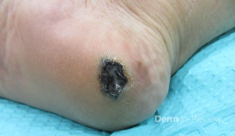
77 year old woman with no personal or family history of skin cancer presented with a lesion on her R heel that she noticed 2 months prior. She had not noticed any growth of the lesion and it was not tender. On physical exam she had a 1.2 x 0.9 cm dark brown/black plaque with hyperkeratosis on the R plantar heel with no lymphadenopathy (Image 1).  On pathology, there was an asymmetric proliferation of atypical melanocytes disposed as nests and single cells present within the epidermis of volar skin and a dermal component identified to 1.2 mm with ulceration. She underwent wide local excision and sentinel lymph node biopsy, which was positive for two lymph nodes with microscopic invasion.

Based on the following information: What is the patient’s pathological tumor stage and overall AJCC8 stage?

A.) T Stage pT2a, AJCC8 Stage IIb
B.) T Stage pT2b, AJCC8 Stage IIIb
C.) T Stage pT2a, AJCC8 Stage IIIb
D.) T Stage pT2b, AJCC8 Stage IIIa
E.) T Stage pT3b, AJCC8 Stage IIIb

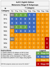
Correct answer: B.) T Stage pT2b, AJCC* Stage IIIb

Based on AJCC8 staging criteria, the pathologic tumor stage would be considered pT2b because the thickness is between 1.0-2.0 mm with ulceration. Answers A and C are incorrect because although the thickness is between 1.0mm and 2.0 mm there is ulceration present making pT2b the correct answer. Answer E is incorrect because the thickness is not >2.0 mm- 4.0 mm, making pT3b incorrect. The nodal disease stage would be N2a because the sentinel lymph node biopsy was positive for two microscopic lymph nodes.  There were not lymph nodes detected clinically so the nodal disease stage would not be N2b. Based on the tumor stage and the nodal stage the AJCC8 stage would be IIIb as shown in the table below. Answer D is not correct because the Stage is IIIb, not IIIa.

According to NCCN guidelines, sentinel lymph node biopsy is generally not recommended for < 0.8 mm.  For tumors with a thickness of 0.8-1.0 mm sentinel lymph node biopsy should be discussed and considered. High risk features to consider for these patients include ulceration, mitoses, and lymphovascular invasion. Sentinel lymph node biopsy should also be discussed and offered to patients with a thickness of > 1.0 mm.

References

  1. Gershenwald JE, Scolyer RA, Hess KR, et al. Melanoma staging: Evidence‐based changes in the American Joint Committee on Cancer eighth edition cancer staging manual. CA: A Cancer Journal for Clinicians. 2017;67(6):472-492. doi:10.3322/caac.21409
  2.  Coit DG, Thompson JA, Algazi A, et al. Melanoma, Version 2.2016, NCCN Clinical Practice Guidelines in Oncology. Journal of the National Comprehensive Cancer Network : JNCCN. 2016;14(4):450-473.

January 2020 Case Study

By 2020 Case Studies

January 2020 Case Study

The patient is a 40 year old female who presents for evaluation of persistent eyelid dermatitis. The eyelid dermatitis has been present for 1 year and has never completely resolved while using topical corticosteroids or calcineurin inhibitors. She describes the rash as itchy but not painful and has not noticed any lesions in other areas. Currently she uses “all natural” personal and cosmetic products including a “fragrance-free” oil on the face. The patient is diagnosed with allergic contact dermatitis of unspecified trigger and patch testing is performed to the North American Standard Tray and Cosmetic Tray (Image 1). A positive patch reaction is noted on the 96 hour read (Image 2). Which of the following allergen did this patient likely react to?

A.) Dimethylol dihydroxyethylene-urea resin
B.) Lanolin
C.) Paraphenylenediamine
D.)Melaleuca alternifolia
E.) Fragrance mix (cinnamic alcohol, cinnamic aldehyde, hydroxycitronellal, amylcinnamaldehyde, gerianiol, eugenol, isoeugenol, oakmoss)

Correct answer: D. Melaleuca alternifolia

Allergic contact dermatitis (ACD) is an inflammatory skin disorder characterized by delayed type IV hypersensitivity. It has a prevalence of approximately 20% and patients with atopic dermatitis may have an increased risk of developing contact dermatitis.1 The eyelids and periorbital area are common locations for ACD and are frequently due to allergens found in cosmetic and personal products including fragrances, formaldehyde-related preservatives, methylisothiazolinone, and cocamidopropyl betaine.1

Patch testing is the best tool to confirm a diagnosis of ACD.2 It involves applying allergens on a patch to the back or upper outer arm of the patient. The patches are removed after 48 hours where an initial read is taken and then a final read is completed at day 4 or day 5. A positive reaction will reveal an erythematous papule or vesicle with edema.2

Melaleuca alternifolia leaf oil is commonly referred to as tea tree oil and is becoming a frequent cause of ACD. M. alternifolia is a part of the North American Contact Dermatitis Group screening patch tray and has a prevalence of 3.5% of positive reactions.3 Clinically, patients have a localized dermatitis depending on where the product is topically applied. The periorbital area is a common location for cosmetic products.3 Patients may present with erythematous patches to eczematous plaques to bullous or erythema multiforme-like reaction.4 When there is a high suspicion of M. alterifolia as a cause of ACD, patients should be patch tested to the 5% formulation in petrolatum along with the patient’s personal products if possible.4 Allergy avoidance and providing the patient with a list of safe products is a necessary part of treatment.

Other answer choices:

Formaldehyde resins like, dimethylol dihydroxyethylene-urea, are commonly used in fabric production to make them wrinkle or water resistant.5 Lanolin contains sterols or fatty acids and is common in many personal and cosmetic topical lotions and creams.5 Paraphenylenediamine (PPD) is used in hair dye and frequently causes ACD of the scalp, eyelids, ears, forehead, and neck.5 There are 2 fragrance mixes found in the North American Contact Dermatitis Group screening tray which covers many fragrances found in perfumes/colognes, oils, cosmetic, personal and household products.5

References

  1. Nassau, S., Fonacier, L. F. (2020). Allergic contact dermatitis. Med Clin N Am, 104, 61-76.
  2. James, W., Elston, D., Treat, J., Neuhaus, I., & Andrews, G. (2020). In Edinburgh (Ed.), Andrews’ diseases of the skin : Clinical dermatology (Thirteenth edition ed.) Elsevier.
  3. de Groot, A. C., & Schmidt, E. (2016). Tea tree oil: Contact allergy and chemical composition. Contact Dermatitis, 75(3), 129-143. doi:10.1111/cod.12591 [doi]
  4. Larson, D., & Jacob, S. E. (2012). Tea tree oil. Dermatitis, 23(1) Retrieved from https://journals.lww.com/dermatitis/Fulltext/2012/01000/Tea_Tree_Oil.10.aspx
  5. Allergen information. (2020). Information posted to https://www.contactderm.org/public-and-patients/allergen-information