
October 2020 Case Study
by Blair Allais, MD
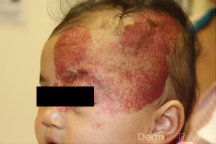
A 2-month-old female infant, born prematurely with a birth weight less than <1000g, presents to dermatology clinic with the following findings. No family history of similar. Physical examination revealed a large hemangioma > 5cm on the face including the scalp.
Which of the following is true regarding her condition?
A.) GNAQ mutations have been identified in these patients
B.) This patient is at risk for high output cardiac failure
C.) Evaluation with head computed tomography (CT) scan typically reveals cortical “tram track” calcifications
D.) This patient is at increased risk for spinal dysraphism
E.) MRI/MRA of the head and neck is indicated to evaluate for anomalies of major cerebral or cervical arteries
Correct answer: E) This patient has PHACE(S) syndrome, and MRI/MRA of the head and neck is indicated to evaluate for cerebrovascular anomalies.
A) False: Somatic mosaic mutations in the GNAQ gene have been described in Sturge-Weber syndrome, which is characterized by capillary malformations in a V1 and V2 > V3 distribution.
B) False: High output cardiac failure is a clinical finding in diffuse neonatal hemangiomatosis (> 5 hemangiomas with organ involvement), kasabach-merritt phenomenon (association with tufted angiomas or kaposiform hemangioendotheliomas) and klippel-trenaunay syndrome (characterized by capillary malformation, venous malformation, and/or lymphatic malformation of a limb).
C) False: Tram-track calcifications are often described on head CTs of patients with sturge-weber syndrome.
D) False: spinal dysraphism is part of the constellation of findings in LUMBAR/SACRAL syndrome.
E) True: This patient has PHACE(S) syndrome, and MRI/MRA of the head and neck is indicated to evaluate for cerebrovascular anomalies.
This infant has PHACE(S) syndrome, which was first described in 19961 for the following spectrum of findings: P, posterior fossa and other structural brain malformations; H, hemangioma; A, arterial anomalies of cervical and cerebral vessels; C, cardiac defects (especially coarctation of the aorta); E, eye anomalies (retinal vascular anomalies, optic nerve hypoplasia); and S, sternal defects and supraumbulical raphe.2 In a study of 108 infants with large facial hemangiomas (>22cm), 31% had PHACES syndrome and 91% of affected individuals had more than one extracutaneous manifestation. The most common findings were cerebrovascular, cardiovascular, and structural brain abnormalities.3 In patients with segmental cervicofacial hemangiomas, PHACE(S) syndrome must be excluded with MRI/MRA of the head and neck, ophthalmologic exam and echocardiogram/cardiac evaluation.
References
Dermatology In Review, Image Companion Figure 3.8.3 PHACES
1.) Frieden IJ, Reese V, Cohen D. PHACE syndrome: the association of posterior fossa abnormalities, arterial anomalies, coarctation of the aorta and cardiac defects, and eye abnormalities. Ach Dermatol 1996; 132:307-11.
2.) Haggstrom AN, Garzon MC. Infantile Hemangiomas. In: Dermatology , Edited by Jean L. Bolognia , Julie V. Schaffer , Lorenzo Cerroni Fourth edition , China: Elsevier, 2018, ISBN 978–0‐7020–6275–9
3.) Haggstrom AN, Garzon MC, Baselga E et al. Risk for PHACE syndrome in infants with large facial hemangiomas. Pediatrics 2010; 126:e418-26