2021 Case Studies – Derm In-Review

December 2021 Case Study

By 2021 Case Studies

December 2021 Case Study

by Azam Qureshi, MD

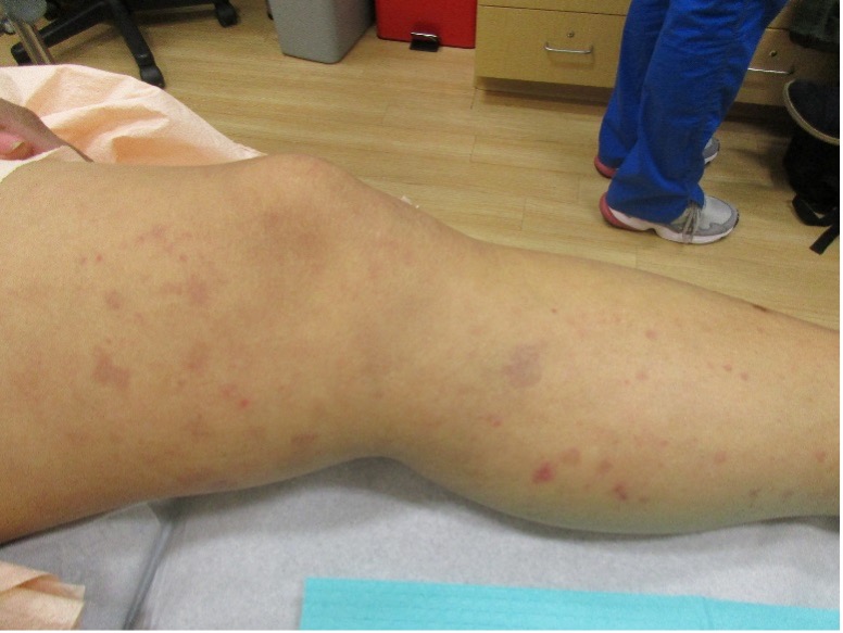
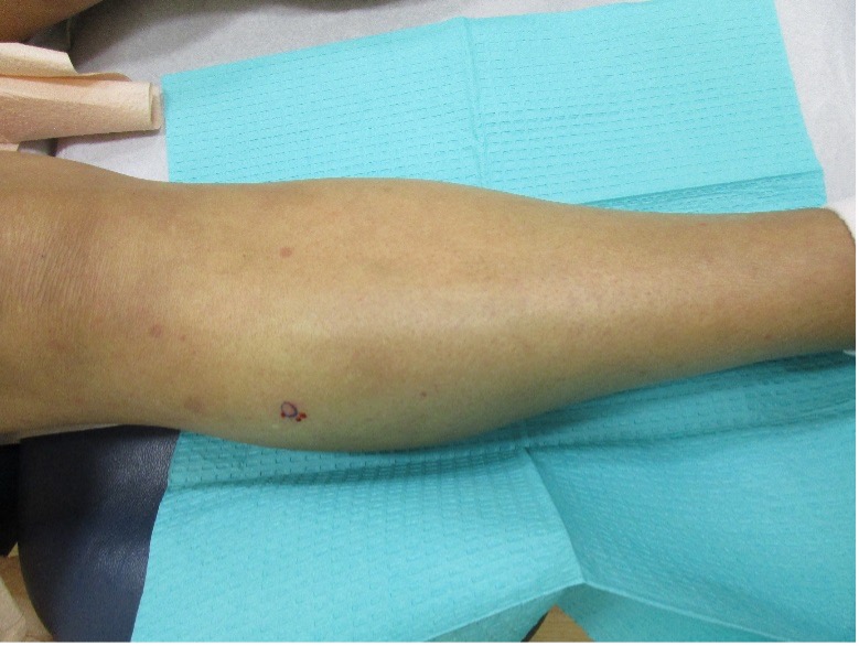
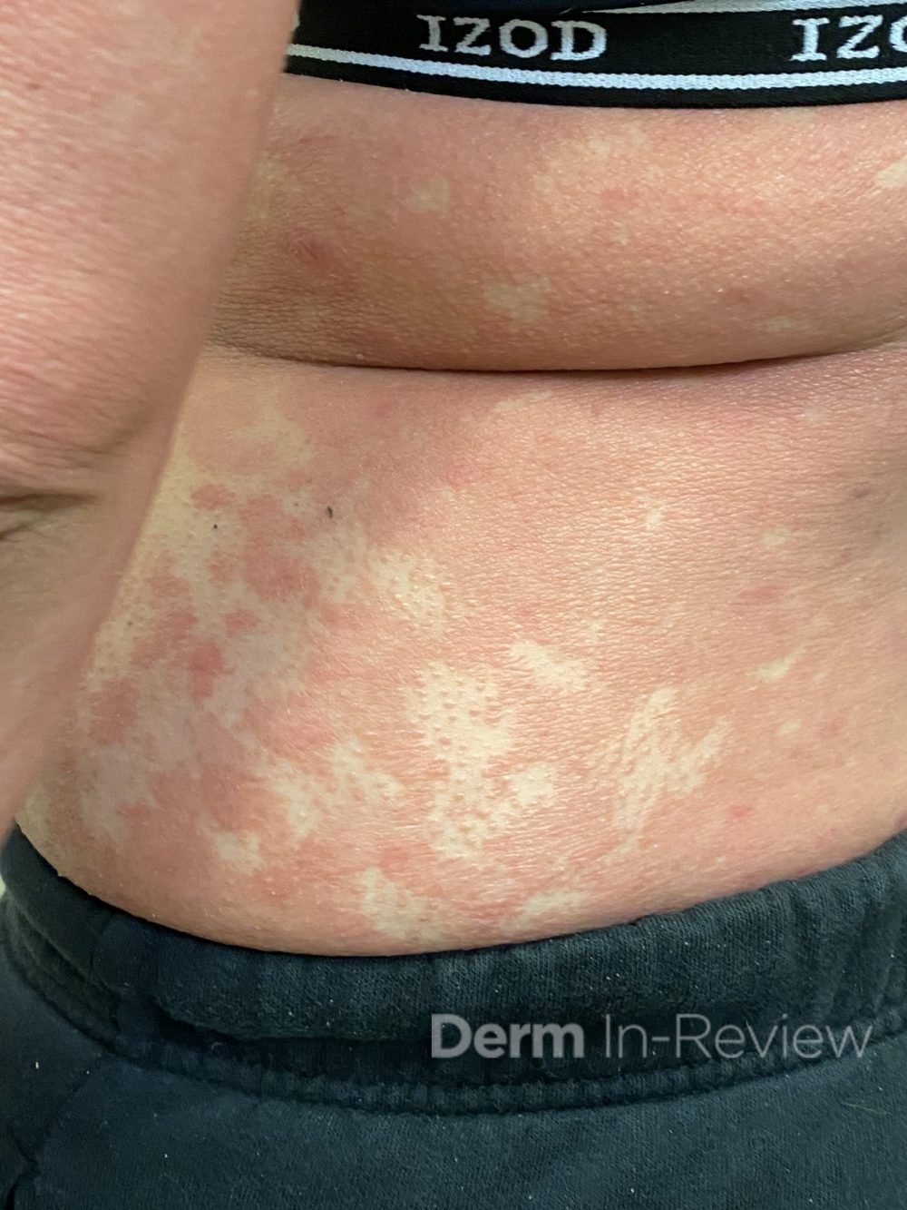
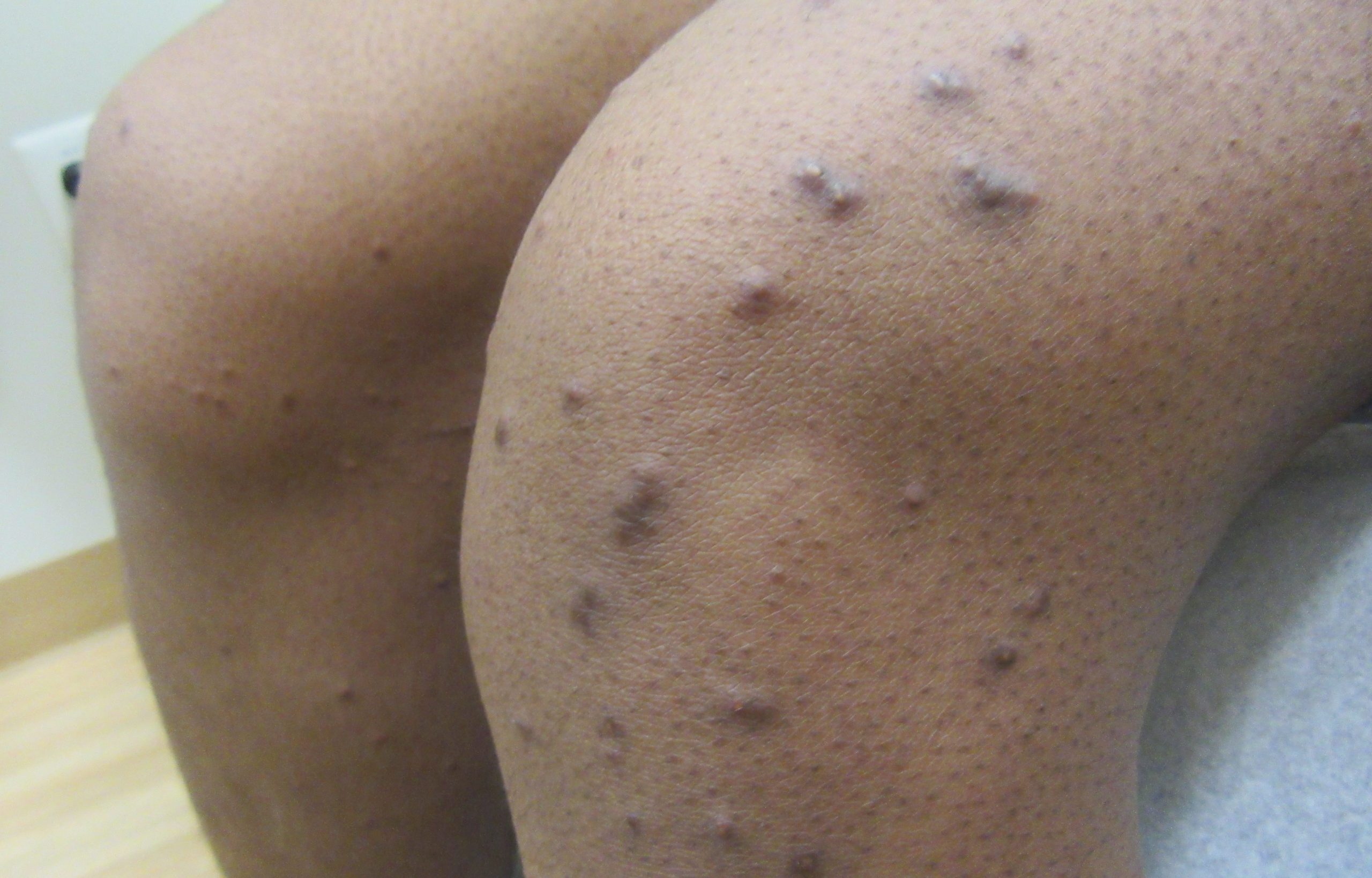
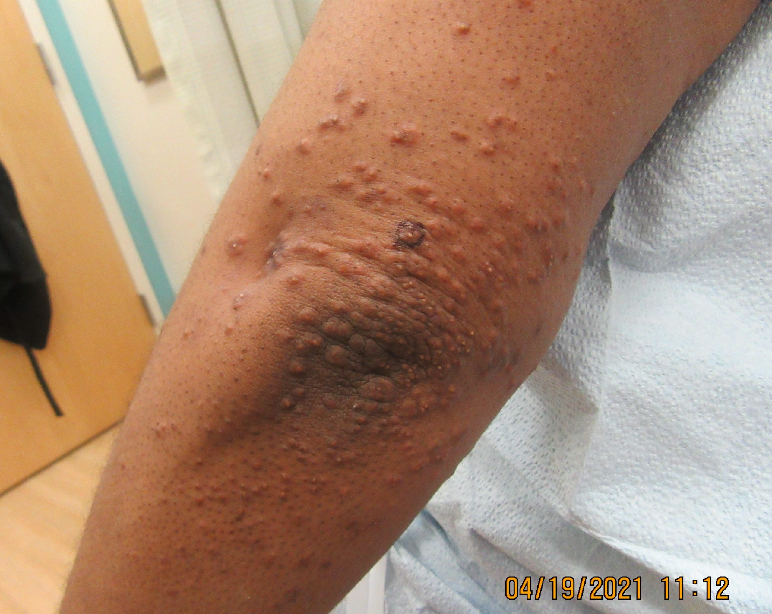
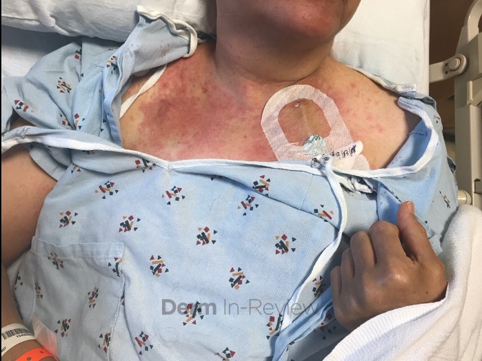
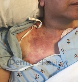
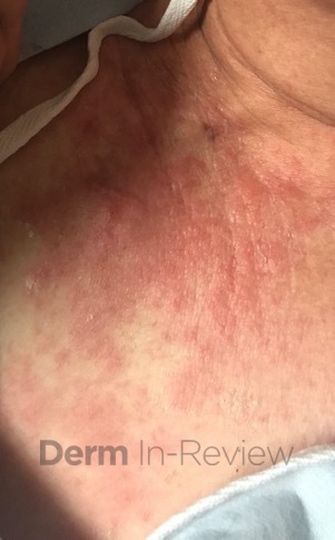
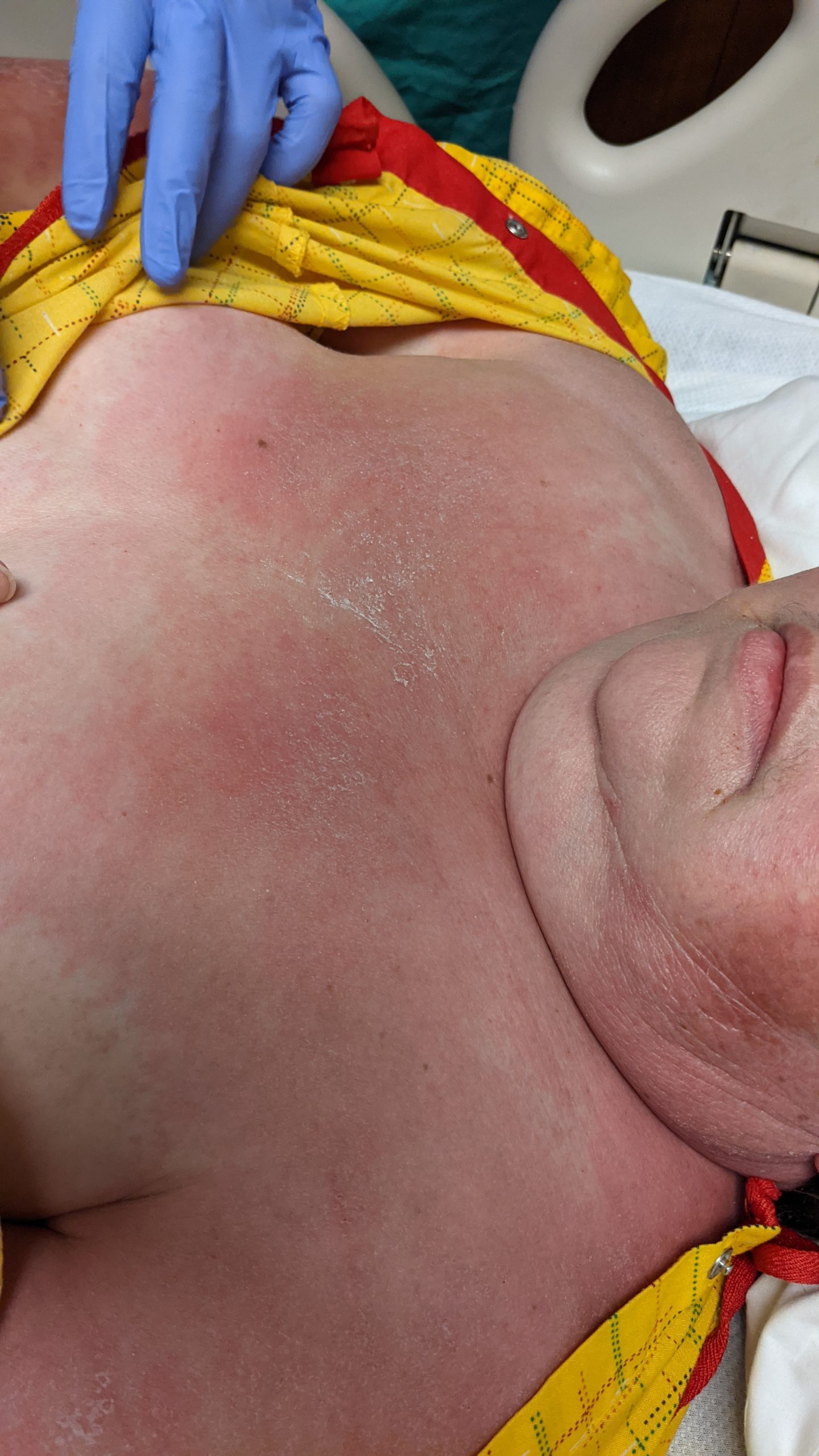
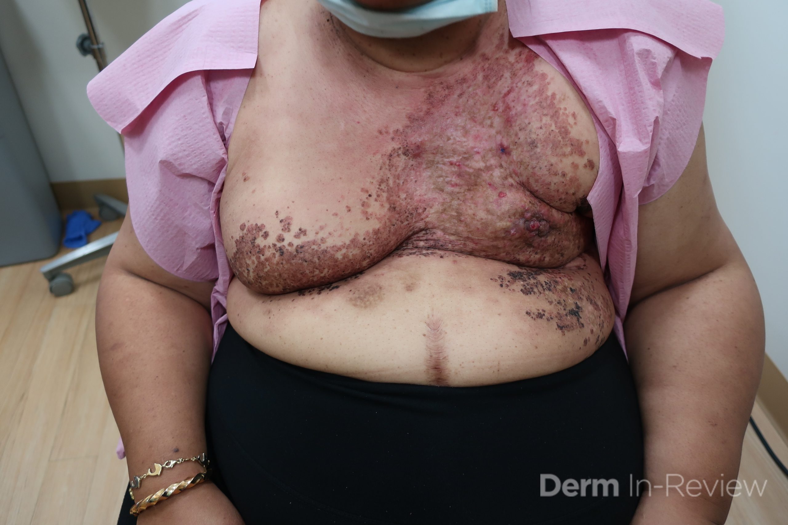
Patient is a 54 year old woman who presents with progressive rash on the anterior torso for 3 months, which she describes as slightly tender bumps. She has history of early stage breast cancer on the left breast treated with lumpectomy and radiation, both completed 9 years ago. She denies any other concerning symptoms.

Biopsy of a representative lesion is most likely to show which of the following?

A.) Dermis infiltrated by atypical neoplasm formed by pleomorphic epithelial cells with hyperchromatic nuclei and distinct nucleoli, disposed in columns and glandular-like structures, amid desmoplastic collagen without associated inflammation, positive immunostaining for HER2, estrogen receptor, and progesterone receptor
B.) Atypical hyperchromatic epithelial cells forming crack-like vascular spaces between collagen bundles, positive immunostaining for CD34, CD31
C.) New vessels wrapping around vascular and adnexal structures, positive immunostaining for HHV-8
D.) Dilation of ectatic superficial vessels, loss of adnexa, papillary dermal pallor, lack of lymphoid band
E.) Acanthosis, pseudoepitheliomatous hyperplasia with suprabasilar crypts of eosinophils

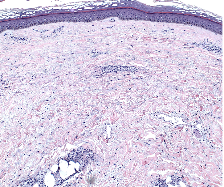
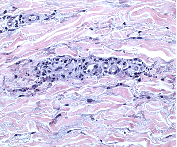
Correct answer: (A) Dermis infiltrated by atypical neoplasm formed by pleomorphic epithelial cells with hyperchromatic nuclei and distinct nucleoli, disposed in columns and glandular-like structures, amid desmoplastic collagen without associated inflammation, positive immunostaining for HER2, estrogen receptor, and progesterone receptor

Explanation:

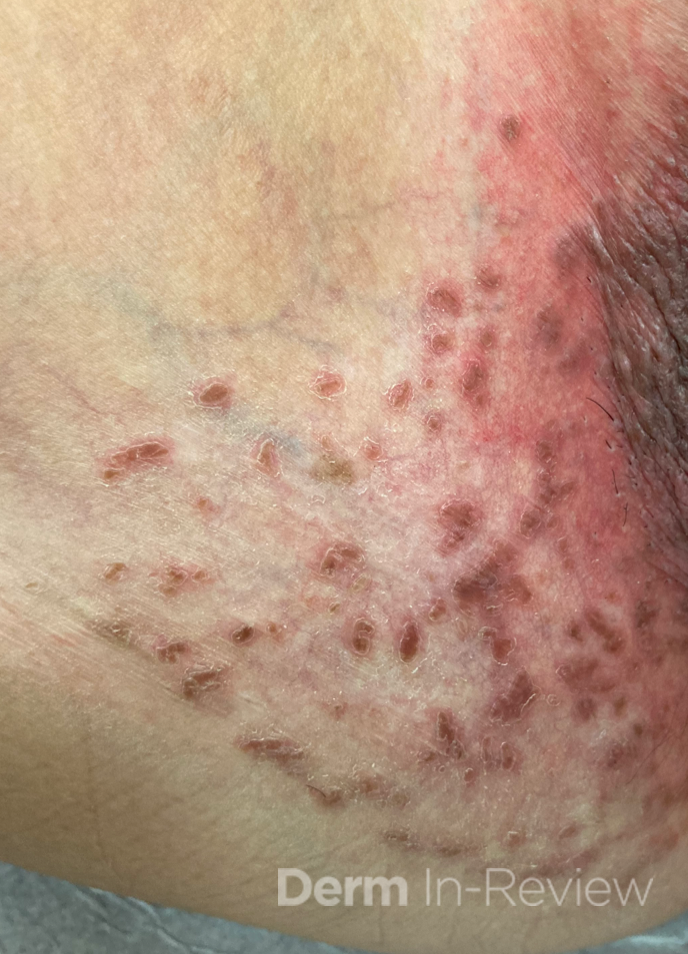
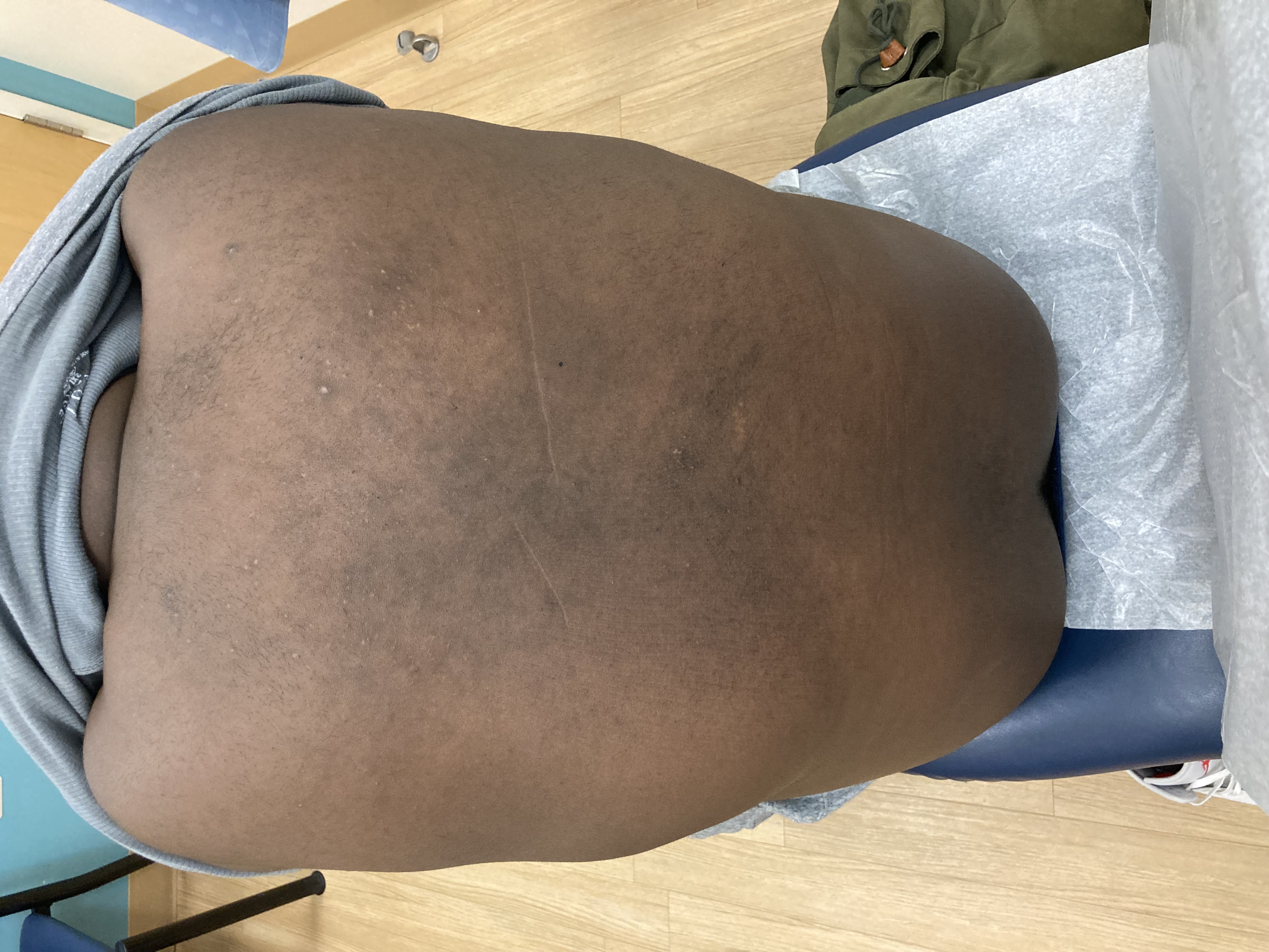
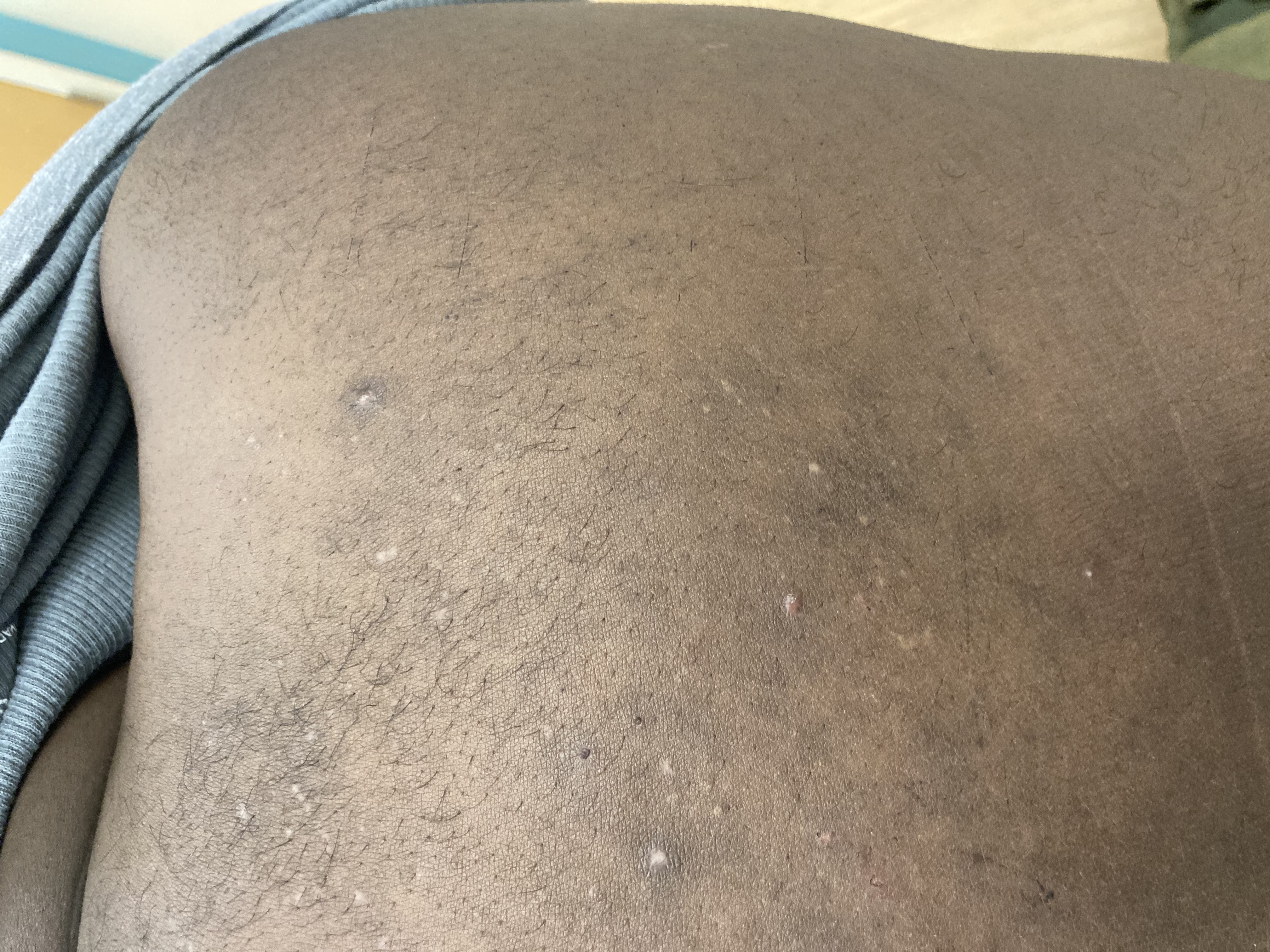
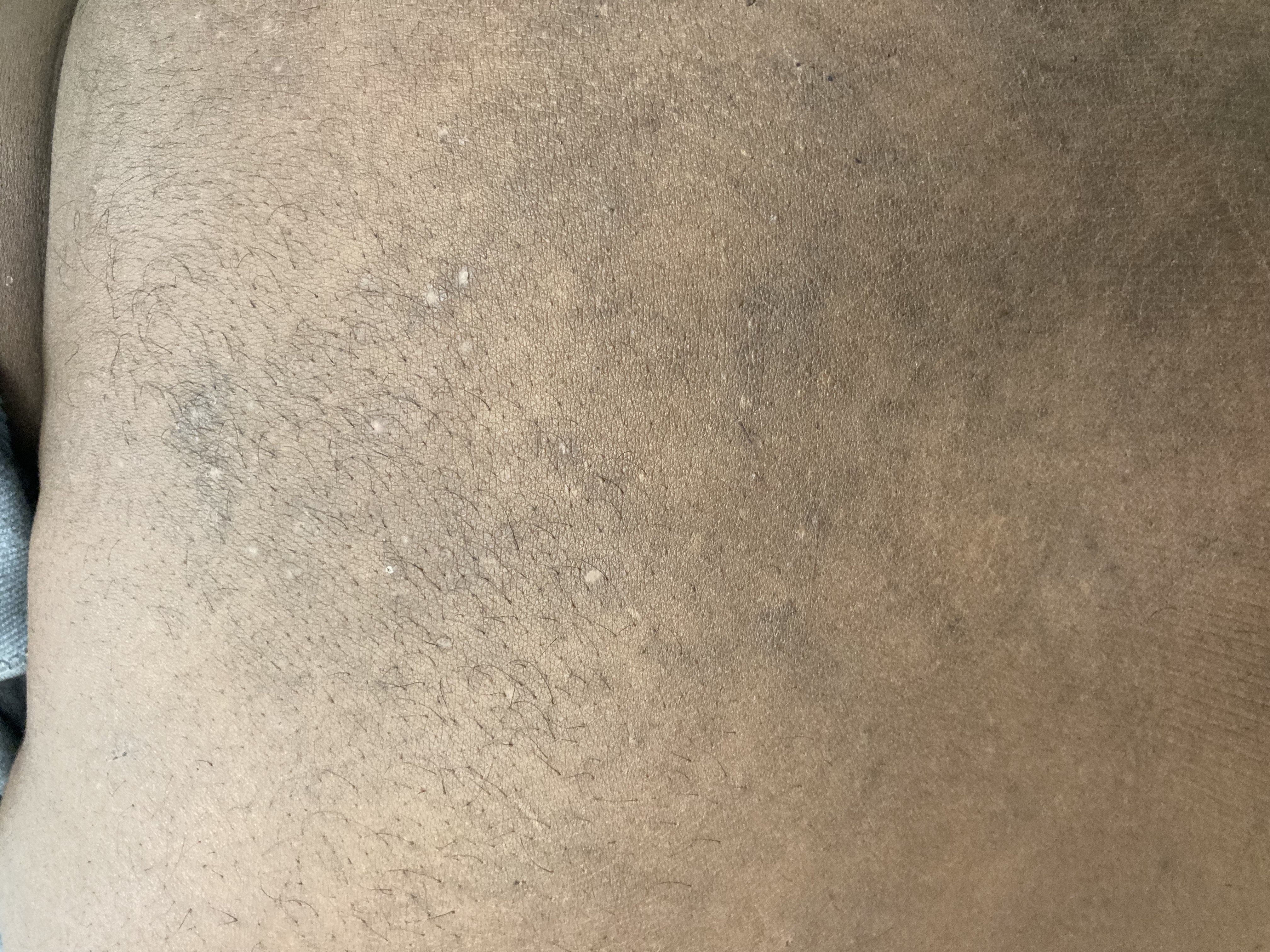
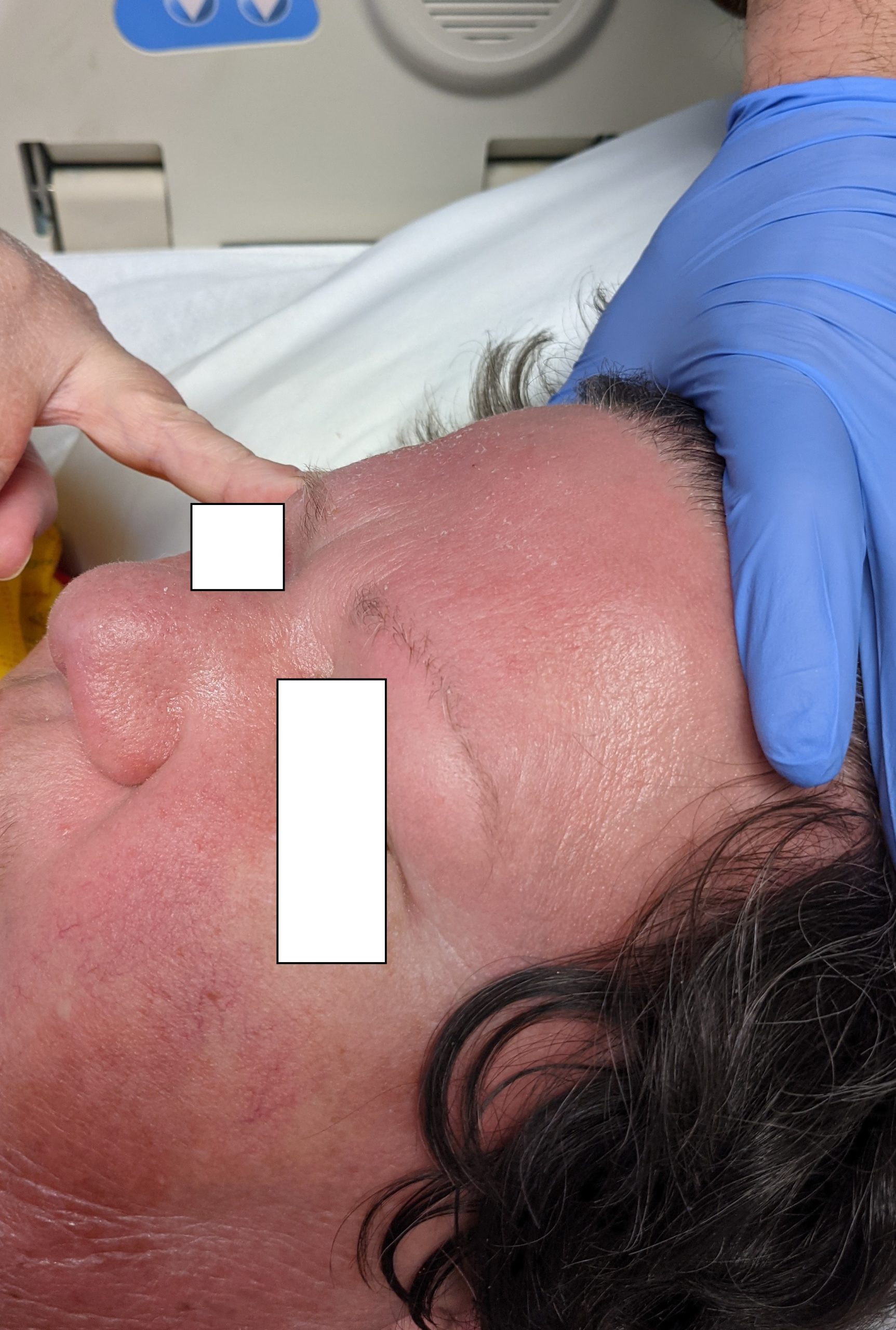
This patient with history of early stage breast cancer who completed surgical and radiation treatment 9 years ago presents with pink to violaceous and hyperpigmented plaques, some studded with pink papules with overlying hemorrhagic crust, predominantly located on the left side of the chest and sprawling out toward the neck and the right chest. Her presentation is most consistent with carcinoma en cuirasse, a presentation of cutaneous metastases most often associated with breast cancer primary. Cutaneous metastases occur in 5-10% of cancer patients and may result from any malignant neoplasm. Prognosis associated with this diagnosis is very poor, as 6 and 12-month mortality rates for these patients are 48% and 64.5%, respectively. Presentations vary, with carcinoma en cuirasse characterized by a diffuse infiltration into the skin, leading to sclerodermoid change. Histopathology may be characterized by a (A) dermis infiltrated by atypical neoplasm formed by pleomorphic epithelial cells with hyperchromatic nuclei and distinct nucleoli, disposed in columns and glandular-like structures, amid desmoplastic collagen without associated inflammation. Positive immunostaining for HER2, estrogen receptor, and progesterone receptor would be suggestive of breast cancer primary. Other presentations of cutaneous metastases include: dermal or subcutaneous nodules, carcinoma erysipeloides, carcinoma telangiectodes, Paget’s disease, alopecia neoplastica, and pyogenic granuloma-like lesions.

Lymphangiosarcoma is high on the differential diagnosis for this patient’s presentation, and may present with non-healing eschars and multiple violaceous nodules. Stewart-Treves syndrome is a rare form of angiosarcoma which arises in the setting of chronic lymphedema, most commonly described in breast cancer patients who have undergone axillary lymph node dissection. Histopathology for this condition may show (B)       atypical hyperchromatic epithelial cells forming crack-like vascular spaces between collagen bundles, with positive immunostaining for CD34, CD31. Patients usually have history of radical mastectomy, as opposed to lumpectomy, and lesions most commonly occur on the ipsilateral upper extremity overlying specific regions of lymphedema, as opposed to widespread lesions occurring over the torso.

Histopathology showing (C) new vessels wrapping around vascular and adnexal structures, with positive immunostaining for HHV-8 would be characteristic of Kaposi sarcoma (KS). There are 4 types of KS, which results directly from human herpesvirus-8 (HHV-8) infection. The classic type usually affects male descendants from the Mediterranean and middle European areas, as well as men in Sub-Saharan Africa, and is not associated with immunodeficiency. Human immunodeficiency virus (HIV)-associated KS is associated with severe immunodeficiency secondary to HIV infection. Endemic KS arises in some parts of Africa, usually in younger patients. Iatrogenic KS results from drug-induced immunosuppression. Presentation classically consists of red to violaceous macules, plaques, or nodules localized anywhere on the skin or mucous membranes, and may even involve the lymph nodes or viscera. There are numerous diverse morphologies reported in the literature, however, including generalized lymphadenopathic, exophytic, disseminated cutaneous and visceral, telangietatic, keloidal, ecchymotic, and localized nodular, plaque, or patch. In this patient who is otherwise well, has no history of immunosuppression, has no known ancestry from any of the aforementioned regions, these widespread tender lesions are more likely representative of cutaneous metastases.

Histopathology showing (D) dilation of ectatic superficial vessels, loss of adnexa, papillary dermal pallor, and lack of lymphoid band would be characteristic of chronic radiation dermatitis. Radiation dermatitis results from direct exposure to radiotherapy delivered by way of external beam ionizing radiation. Acute radiation dermatitis may present as erythema, desquamation, skin necrosis, and ulceration in areas of skin that have been irradiated. Chronic radiation dermatitis may occur up to 10 years or more after the beginning of radiotherapy and involves telangiectasia, fragile skin, and loss of follicular structures. In addition to the current patient’s distinctive morphology suggestive of cutaneous metastases, her radiation was 9 years ago and her eruption is very widespread, making radiation dermatitis much less likely.

Histopathology showing (E) acanthosis, pseudoepitheliomatous hyperplasia with suprabasilar crypts of eosinophils would be suggestive of pemphigus vegetans. Pemphigus vegetans is a localized form of pemphigus vulgaris. Early lesions may present as flaccid pustules, leading to erosions and vegetative and papillomatous plaques usually involving intertriginous areas, scalp, and face. There are 2 predominant subtypes, with the Hallopeau type consisting of more benign disease with longer periods of remission, and the Neumann type involving more disease relapses and requiring higher doses of corticosteroids. Similar to other forms of pemphigus vulgaris, patients have autoantibodies to desmoglein 3 predominantly. This current patient’s lesion morphology and areas of involvement make pemphigus vegetans an unlikely diagnosis.

References

  1. Bolognia JL, Schaffer JV, Duncan KO, Ko CJ. Dermatology essentials E-book. Elsevier Health Sciences; 2014 Feb 26.
  2. Elston D, Ferringer T, Ko CJ, Peckham S, High WA, DiCaudo DJ. Dermatopathology E-Book. Elsevier Health Sciences; 2013 Oct 16.
  3. Schoenlaub, P., et al. “Survival after cutaneous metastasis: a study of 200 cases.” Annales de Dermatologie et de Venereologie. Vol. 128. No. 12. 2001.
  4. Oliveira GM, Zachetti DB, Barros HR, Tiengo A, Romiti N. Breast carcinoma en Cuirasse-case report. Anais brasileiros de dermatologia. 2013 Aug;88(4):608-10.
  5. Mahore SD, Bothale KA, Patrikar AD, Joshi AM. Carcinoma en cuirasse: A rare presentation of breast cancer. Indian Journal of Pathology and Microbiology. 2010 Apr 1;53(2):351.
  6. Sharma A, Schwartz RA. Stewart-Treves syndrome: pathogenesis and management. Journal of the American Academy of Dermatology. 2012 Dec 1;67(6):1342-8.
  7. Etemad SA, Dewan AK. Kaposi sarcoma updates. Dermatologic clinics. 2019 Oct 1;37(4):505-17.
  8. Hegedus F, Mathew LM, Schwartz RA. Radiation dermatitis: an overview. International journal of dermatology. 2017 Sep;56(9):909-14.
  9. Razzaque Ahmed A, Blose DA. Pemphigus vegetans: Neumann type and Hallopeau type. International journal of dermatology. 1984 Mar;23(2):135-41.

November 2021 Case Study

By 2021 Case Studies

November 2021 Case Study

by Dillon Nussbaum, MD

A 31-year-old female with no past medical history presents for evaluation of the pictured lesions of the right upper thigh. The lesions have been present since birth and occasionally pruritic. She was previously treated unsuccessfully with electrodessication, topical sulfur, and combination betamethasone-gentamycin in another country. She denies any systemic symptoms and has no personal or family history of skin conditions other than the pictured lesions.

Immunohistochemical staining of these lesions would most likely be positive for all of the following EXCEPT:

A.) LYVE-1
B.) S-100
C.) Prox1
D.) Podoplanin
E.) VEGFR-3

Correct answer: B

Explanation:

The history and image provided are most consistent with a microcystic lymphatic malformation, previously known as lymphangioma circumscriptum. Microcystic lymphatic malformations are usually a result of congenital abnormalities in the development of superficial lymphatic channels, but they can be acquired following trauma, radiation, or a malignancy. They typically appear as grouped clear, yellow, or red appearing papules and/or vesicles resembling frog spawn.

Histopathology of microcystic lymphatic malformations reveals enlarged and abnormal appearing lymphatic vessels with smooth muscle cells lining thin walled endothelium. The lymphatic endothelium can be identified by immunohistochemical staining for LYVE1, Prox1, Podoplanin, and VEGFR-3.1

LYVE-1, Prox1, podoplanin (D2-40), and VEGFR-3 are all immunohistochemical stains for lymphatic tissue and therefore will be positive when staining the pictured lesions. These markers are also useful for identifying angiosarcoma, kaposi sarcoma, lymphangiosarcoma, and mesothelioma among other vascular and lymphatic based conditions or tumors. However, the remaining answer, S-100, is generally positive in in melanoma, nerve sheath tumors, schwannomas, clear cell sarcoma of soft tissue, various histiocytic tumors, glial tumors and myoepithelial tumors.2,3

There are no standard treatment guidelines for microcystic lymphatic malformations. Depending on location and presentation, treatment can consist of a combination of surgical excision, sclerotherapy, laser therapy, hyfrecation, cryotherapy, and topical sirolimus. Macrocystic lymphatic malformations are seen commonly in turner syndrome as cystic hygromas on the neck; microcystic lymphatic malformations are less commonly associated with syndromic pictures.4

References

  1. Davis M, van der Hilst J. Mimickers of urticaria: Urticarial vasculitis and autoinflammatory diseases. J Allergy Clin Immunol Pract. 2018. 6(4):1162-1170.
  2. Bolognia, Jean L., MD; Schaffer, Julie V., MD; Cerroni, Lorenzo, MD. Dermatology, Fourth Edition. 2018.
  3. Gu SL, Jorizzo JL. Urticarial vasculitis. Int J Womens Dermatol. 2021 Jan 29;7(3):290-297. doi: 10.1016/j.ijwd.2021.01.021. PMID: 34222586; PMCID: PMC8243153.
  4. Magro C, Nuovo G, Mulvey J, Laurence J, Harp J, Crowson AN. The skin as a critical window in unveiling the pathophysiologic principles of COVID-19. Clin Dermatol. 2021 Jul 22. doi: 10.1016/j.clindermatol.2021.07.001. Epub ahead of print. PMCID: PMC8298003.
  5. Jara LJ, Navarro C, Medina G, Vera-Lastra O, Saavedra MA. Hypocomplementemic urticarial vasculitis syndrome. Curr Rheumatol Rep. 2009 Dec;11(6):410-5. doi: 10.1007/s11926-009-0060-y. PMID: 19922730.

October 2021 Case Study

By 2021 Case Studies

October 2021 Case Study

by Emily Murphy, MD, Karl Saardi, MD

A 38-year-old male with a past medical history of HIV presented with an intermittent, itchy rash for 1.5 years. The rash first occurred on his upper arms but now predominantly affects his upper back. He has not tried any therapies (figure 1). Of note, he was off anti-retroviral therapy (ART) for several years resulting in a low CD4 count of 15. He restarted ART three months prior to presentation but this did not affect the course of his rash. One of the pustules was de-roofed and examined after application of potassium hydroxide (figure 2).

Which of the following statements is true about this eruption?

A.) This disease is more likely in patients with HIV regardless of CD4 count.
B.) While potassium hydroxide staining of skin scrapings may help with the diagnosis, the pathogen is only rarely seen on microscopy.
C.) This eruption can be seen as part of immune inflammatory reconstitution syndrome.
D.) Compared to this eruption, papular pruritic eruption of HIV has a similar clinical appearance and distribution on the face, neck, and trunk.
E.) One possible treatment is a two week course of Ivermectin.

Correct Answer: C

Explanation/Literature review:

The demodex mite is a normal colonizer of the skin; 69% of patients aged 31 to 50 years old are colonized and 95% of patients over 71 years old are colonized.1 Demodex folliculitis occurs when there is inflammation of pilosebaceous units infested with mites. Bacterial antigens on the mites trigger a lymphocytic infiltrate, producing the erythematous perifollicular papules and pustules that are seen with this disease.1 The disease tends to occur on the face and neck but can be more diffuse.1 A skin scraping or de-roofing of a pustule and staining with potassium hydroxide will usually reveal the demodex mite on microscopy (answer B).1,2 There are two types of mites. Demodex folliculorium is the larger mite that is often present on the face, clustered in groups of 10-15 mites on the follicular infundibulum. Demodex brevis is the smaller mite that often exists on the trunk as a solitary mite in the sebaceous gland.1

Demodex folliculitis is more common in immunosuppressed patients, especially patients with HIV who have a CD4 count less than 200 (answer A).1,2 Immunosuppressed patients may have more diffuse disease involving the sebaceous areas of the face, upper chest, and interscapular back.1,2 While demodex folliculitis is often seen with low CD4 counts, it can also paradoxically occur with immune reconstitution inflammatory syndrome, when patients have an increasing CD4 count after starting ART (answer C).2 This phenomenon may be due to uncontrolled replication of the mites or a local influx of T cells with immune reconstitution.2 Demodex folliculitis can be seen with other immunosuppressed patients, including patients with leukemia who undergo stem cell transplant, where this eruption can mimic acute graft versus host disease.3

Beyond demodex folliculitis, other diseases should be considered when a patient with HIV presents with pruritic papules and pustules, including papular pruritic eruption of HIV, immunosuppression-associated eosinophilic folliculitis, and pityrosporum folliculitis. These four eruptions are morphologically similar but the distribution of papular pruritic eruption of HIV differs, often presenting on the extremities, rather than the face, neck, and upper trunk in the other three diseases (answer D).1,3,4

Demodex folliculitis can be treated topically or orally, with the goal of reducing the demodex mite infestation. Response to therapy confirms pathogenicity of the mite.1 Topical treatments include sulfur products like sodium sulfacetamide 10% lotion or shampoo, ivermectin 1% cream, permethrin 5% cream, or metronidazole 1% cream/gel.1,5 More diffuse or recalcitrant disease should be treated orally with a single dose of ivermectin 200 micrograms/kilogram (12-15 milligrams) (answer E).1,5 Initial oral therapy can be followed by intermittent topical therapy to minimize the long-term population of demodex mites.

References

  1. Elston CA, Elston DM. Demodex mites. Clin Dermatol. 2014;32(6):739-743. doi:10.1016/j.clindermatol.2014.02.012
  2. Delfos NM, Collen AFS, Kroon FP. Demodex folliculitis: a skin manifestation of immune reconstitution disease. AIDS. 2004;18(4):701-702. doi:10.1097/00002030-200403050-00019
  3. Cotliar J, Frankfurt O. Demodex folliculitis mimicking acute graft-vs-host disease. JAMA Dermatol. 2013;149(12):1407-1409. doi:10.1001/jamadermatol.2013.5891
  4. Motswaledi MH, Visser W. The spectrum of HIV-associated infective and inflammatory dermatoses in pigmented skin. Dermatol Clin. 2014;32(2):211-225. doi:10.1016/j.det.2013.12.006
  5. Jacob S, VanDaele MA, Brown JN. Treatment of Demodex-associated inflammatory skin conditions: A systematic review. Dermatol Ther. 2019;32(6):e13103. doi:10.1111/dth.13103

September 2021 Case Study

By 2021 Case Studies

September 2021 Case Study

by Kamaria Nelson, MD

A 51-year-old female with a past medical history of autoimmune hepatitis and mixed connective tissue disease, presents with a recurrent rash on the legs and buttocks for the past 3 months. Patient describes the rash as red and pruritic. The rash occurs every 8 days, lasts for roughly 2-3 days, and resolves leaving “dark spots.” She is currently not using any medication for the rash. Physical exam revealed purple to brown macules and red, thin small plaques on the bilateral thighs and legs (Images 1-2). No lesions noted on the buttocks. A punch biopsy was performed (Images 3-4).

Based upon the patient history, clinical examination, and histopathologic findings, which laboratory finding is the most important prognostic feature in this skin condition?

A.) Positive ANA
B.) Elevated ESR
C.) Anti-C1q precipitin
D.) Low serum C3/4 levels
E.) Low C1q levels

Urticarial vasculitis (UV) is characterized by persistent urticarial lesions lasting for longer than 24 hours with histopathologic findings of leukocytoclastic vasculitis (LCV).  Urticarial lesions are present at onset and often resolve with dusky/hyperpigmented patches and plaques after resolution. UV has an incidence of 5 cases/million/year and prevalence of about 5%. It has a slight female predominance and peaks in the 4th and 6th decades of life. Approximately 80% of cases have a benign course and resolve within 3 years. UV has a similar pathogenesis of typical cutaneous small vessel vasculitis except it is thought to be a type III hypersensitivity reaction with antibody complexes that activate complement which triggers mast cell release of inflammatory mediators like TNF-α. This leads to increase of ICAM on mast cells and E-selectin on endothelial cells. There are several precipitating factors of UV including autoimmune connective tissue diseases, infections (i.e. hepatitis B and C), medications (i.e. methotrexate, NSAIDs), hematologic malignancies and rarely solid organ malignancies.  Recently, studies have shown UV to be associated with COVID-19 infection.

Work-up for UV includes skin biopsy with direct immunofluorescence (DIF) which will reveal leukocytoclasis, fibrinoid deposits around blood vessels, neutrophilic perivascular infiltrate, extravasation of RBCs, and injury and swelling of endothelial cells. DIF will show immunoglobulin and complement deposition around blood vessels. Age-appropriate screening for malignancies is also warranted. Laboratory studies include CBC, CMP, ESR, hepatitis studies, urinalysis, complement studies, C1q levels, and anti-C1q antibody assays. Autoimmune work-up may include ANA, anti-RNP, anti-Smith, Anti-SS-A/B, Anti-Scl-70, Anti-Jo-1, and Anti-Centromere antibodies. The most important prognostic feature is the presence of hypocomplementemia (answer D). Other laboratory findings include elevated ESR (answer B), positive ANA (answer A), presence of anti-C1q precipitin (answer C), and depressed C1q levels (answer E).

Patients with normocomplementemia usually have skin-limited disease and patients with hypocomplementemia have an increased risk of systemic manifestations. Hypocomplementemic urticarial vasculitis syndrome (HUVS) is a more severe form of UV and is associated with systemic symptoms. Major criteria include urticaria for 6 months and hypocomplementemia. Minor criteria include vasculitis on biopsy, arthralgia, uveitis, glomerulonephritis, recurrent abdominal pain, and positive C1q precipitin test. In order to diagnose HUVS, patients must meet both major criteria and 2 or more minor criteria.

Treatments for UV is the same for normo and hypocomplementemia. 1st line therapies include antihistamines, oral corticosteroids, indomethacin and dapsone. Other therapies include colchicine, hydroxychloroquine, methotrexate, dapsone, azathioprine, and mycophenolate mofetil. Rituximab, IVIG, omalizumab, and interleukin-1 inhibitors may be used for recalcitrant hypocomplementemic UV. Treatment is often difficult because of the lack of large randomized controlled trials and there are currently no drugs FDA-approved for use in UV. Symptoms may improve after treatment of underlying conditions in affected patients.

References

  1. Davis M, van der Hilst J. Mimickers of urticaria: Urticarial vasculitis and autoinflammatory diseases. J Allergy Clin Immunol Pract. 2018. 6(4):1162-1170.
  2. Bolognia, Jean L., MD; Schaffer, Julie V., MD; Cerroni, Lorenzo, MD. Dermatology, Fourth Edition. 2018.
  3. Gu SL, Jorizzo JL. Urticarial vasculitis. Int J Womens Dermatol. 2021 Jan 29;7(3):290-297. doi: 10.1016/j.ijwd.2021.01.021. PMID: 34222586; PMCID: PMC8243153.
  4. Magro C, Nuovo G, Mulvey J, Laurence J, Harp J, Crowson AN. The skin as a critical window in unveiling the pathophysiologic principles of COVID-19. Clin Dermatol. 2021 Jul 22. doi: 10.1016/j.clindermatol.2021.07.001. Epub ahead of print. PMCID: PMC8298003.
  5. Jara LJ, Navarro C, Medina G, Vera-Lastra O, Saavedra MA. Hypocomplementemic urticarial vasculitis syndrome. Curr Rheumatol Rep. 2009 Dec;11(6):410-5. doi: 10.1007/s11926-009-0060-y. PMID: 19922730.

August 2021 Case Study

By 2021 Case Studies

August 2021 Case Study

by Jessica Kalen, MD

A 37-year-old female with no past medical history presents for evaluation of the pictured rash over the body. Initially began 6 weeks ago with significant associated pruritus. Prior to presentation was treated with methylprednisolone taper and triamcinolone 0.1% ointment. Additional clinical findings include a keratoderma of the bilateral hands and yellowing of fingernails with subungual debris. She denies joint pain or stiffness, but does report malaise and chills. She has no personal or family history of skin conditions.

Based on the clinical presentation, what are the anticipated histopathological findings for this condition?

A.) Regular acanthosis with neutrophilic debris and parakeratosis in stratum corneum
B.) Shoulder parakeratosis and neutrophilic debris focused around follicular ostia
C.) Alternating vertical and horizontal orthokeratosis and parakeratosis, acanthosis, and mild spongiosis
D.) Subacute spongiosis with extravasation and transepidermal elimination of red blood cells
E.) Lichenoid interface dermatitis with hypergranulosis and dyskeratosis

Correct answer: C

Explanation

Based on the clinical presentation, the anticipated histopathological findings would be most consistent with pityriasis rubra pilaris (PRP). This condition presents with widespread waxy, orange plaques and islands of sparing as well as prominent follicular hyperkeratosis, often compared to a nutmeg grater.1 Onset occurs over the course of several weeks with an, often, rapid progression to erythroderma.1 PRP is classified into six subtypes based on age, duration, and clinical appearance. Subtypes include classic adult, atypical adult, classic juvenile, circumscribed juvenile, atypical juveline, and HIV-associated.1

Histopathologically, the classic finding of PRP is alternating orthokeratosis and parakeratosis both vertically and linearly.2 This is often compared to a “checkboard”. Additionally, there is follicular plugging with shoulder parakeratosis, hypergranulosis, mild spongiosis, and sparse lymphohistiocyctic infiltrate.1

Standardized treatment guidelines of PRP remain to be established. However, several systemic medications have been empirically utilized to treat this condition. PRP has been successfully treated with oral retinoids, methotrexate, cyclosporine, and azathioprine.1 Now with the advent of biologics, PRP has also shown good response to TNF-alpha inhibitors, guselkumab, ixekizumab, secukinumab.1,3,4

The remaining answers do not describe the histopathological findings of PRP. Choice A describes findings of psoriasis, choice B describes seborrheic dermatitis, choice D describes pityriasis rosea, and choice E describes lichen planus.

References

  1. Bolognia JL, Schaffer JV, Cerroni L. Dermatology. 4th Philadelphia. Elsevier Limited.
  2. Elston DM, et al. Dermatopathology. 4th 2019. Elsevier.
  3. Pilz AC, et al. Treatment of pityriasis rubra pilaris with guselkumab. JAMA Dermatol. Dec 2019;155(12):1424-1426.
  4. Haynes D, et al. Evaluation of ixekizumab treatment for patients with pityriasis rubra pilaris: a single-arm trial. JAMA Dermatol. 2020;156(6):668-675.

July 2021 Case Study

By 2021 Case Studies

July 2021 Case Study

by Azam Qureshi, MD

Patient is a 37 year old woman who presents with rash on the extremities for 3 months. The asymptomatic bumps started on the right elbow and subsequently spread to the left elbow and bilateral knees. She denies any other concerning symptoms.

Which of the following other laboratory findings is also most likely to be present?

A.) Elevated hemoglobin a1c
B.) Low thyroid-stimulating hormone
C.) Elevated pancreatic lipase
D.) Elevated serum sodium & osmolality along with decreased urine osmolality & specific gravity
E.) Monoclonal spike on serum protein electrophoresis

Answer: (A) Elevated hemoglobin a1c

This patient presents with crops of pink to yellow papules, some with an erythematous halo, about 1 to 5 mm in diameter distributed on the extensor surfaces of the extremities, most consistent with eruptive xanthomas.

Elevated hemoglobin a1c levels (A) above 6.5% are suggestive of diabetes mellitus, which is associated with impaired insulin activity. Deficiency or resistance to insulin both causes a decrease in lipoprotein lipase activity while thereby also contributing to hepatic overproduction of triglyceride-rich lipoproteins, both effectively serving to contribute to pathogenesis of eruptive xanthomas.1 Lipoprotein lipase is bound to capillary endothelium and is involved in both exogenous and endogenous pathways of triglyceride (TG) and cholesterol circulation. This enzyme releases free fatty acids to the peripheral tissues by catalyzing the hydrolysis of core TGs in circulating chylomicrons and VLDL molecules by way of complex interactions involving hormones, including insulin, and apoproteins, including apo C-II.1 The etiology of eruptive xanthomas is intricately related to deficiency of lipoprotein lipase activity and hepatic overproduction of TG-rich lipoproteins, both of which can result or be worsened by impaired insulin activity.1 Other environmental factors playing important roles in the appearance of eruptive xanthomas include: hypothyroidism, alcohol abuse, estrogen replacement therapy, systemic retinoids, anti-retroviral therapy, olanzapine, and azacitidine.1,2

Low thyroid-stimulating hormone (B) is suggestive of hyperthyroidism. Although hypothyroidism has been shown to be a contributing factor to the presentation of eruptive xanthomas, hyperthyroidism is not a common trigger.1,2 Common dermatologic findings associated with hyperthyroidism include pretibial myxedema, hyperhidrosis, flushing, diffuse hair thinning, onycholysis, and Plummer’s nail.3

When occurring in the context of hypertriglyceridemia, TG levels in patients with eruptive xanthomas often exceed 3,000 to 4,000.1 Acute pancreatitis, associated with elevated pancreatic lipase levels (C), occurs at a significantly increased risk in patients with very high TG levels, and has been shown to occur in approximately 10-20% in patients with TG greater than 2,000.4 Although eruptive xanthomas may be an important warning sign that may herald the onset of acute pancreatitis, the laboratory finding of elevated lipase in a patient with no other concerning symptoms would be less likely than a finding of elevated hemoglobin a1c, as diabetes mellitus is a common precipitating factor for eruptive xanthomas.

Diabetes insipidus is characterized by the laboratory findings of elevated serum sodium & osmolality along with decreased urine osmolality & specific gravity (D), and has been associated with both xanthoma disseminatum and Erdheim-Chester disease, both non-Langerhans cell histiocytoses (LCH).1 Xanthoma disseminatum is a normolipemic type of non-LCH which presents with a triad including cutaneous xanthomas, mucosal xanthomas, and diabetes insipidus.1 Xanthomas in this condition more commonly present with symmetric flexural and intertriginous involvement, however. Erdheim-Chester disease is another non-LCH which may also present with diabetes insipidus, along with fever, multiorgan involvement, other neurologic symptoms, and bone lesions and fractures.1 Although this condition may present with red to brown to yellow indurated plaques and nodules, skin involvement is infrequent in this rare condition. Both of these conditions are also much less likely in this patient with no other concerning symptoms.

Monoclonal spike on serum protein electrophoresis (E) is indicative of plasma cell dyscrasia, which has been associated with aforementioned xanthoma disseminatum, normolipemic plane xanthomas, along with number of other dermatologic conditions including Sweet’s syndrome, AL amyloidosis, necrobiotic xanthogranuloma, scleredema, scleromyxedema, IgA pemphigus, subcorneal pustular dermatosis, pyoderma gangrenosum, erythema elevatum diutinum, POEMS syndrome, Schnitzler’s syndrome, cryoglobulinemia, and Waldenstrom’s macroglobulinemia.1,5-7 Plane xanthomas classically present around the periorbital region, lateral neck, upper trunk, flexures, interdigital spaces, and palmar creases. The patient’s presentation is more consistent with eruptive xanthomas.

References

  1. Bolognia JL, Schaffer JV, Duncan KO, Ko CJ. Dermatology essentials E-book. Elsevier Health Sciences; 2014 Feb 26.
  2. Babade, M., Prodanovic, E., Mostow, E. Eruptive Xanthoma Associated with Diabetic Ketoacidosis: Lessons from a Case. Practical Dermatology. May 2006.
  3. Lause M, Kamboj A, Faith EF. Dermatologic manifestations of endocrine disorders. Translational pediatrics. 2017 Oct;6(4):300.
  4. Scherer J, Singh V, Pitchumoni CS, Yadav D. Issues in hypertriglyceridemic pancreatitis-an update. Journal of clinical gastroenterology. 2014 Mar;48(3):195.
  5. Iglesias-Girard L, Roy SF, Chapdelaine H, Désy D, Bouffard D, Funaro D. Disseminated Xanthosiderohistiocytosis With Monoclonal Gammopathy—A Rare Form of Xanthoma Disseminatum. JAMA dermatology. 2020 Nov 1;156(11):1270-2.
  6. Cohen YK, Elpern DJ. Diffuse normolipemic plane xanthoma associated with monoclonal gammopathy. Dermatology practical & conceptual. 2015 Oct;5(4):65.
  7. Daoud MS, Lust JA, Kyle RA, Pittelkow MR. Monoclonal gammopathies and associated skin disorders. Journal of the American Academy of Dermatology. 1999 Apr 1;40(4):507-35.

June 2021 Case Study

By 2021 Case Studies

June 2021 Case Study

by Adrianna Gonzalez, MD

A 63-year-old woman was admitted following a hematopoietic stem cell transplant (HSCT) for the treatment of multiple myeloma. Eleven days after her transplant, she developed a morbilliform eruption on her back that subsequently spread to her chest, abdomen and upper arms (Figure 1a-b). The rash was initially pruritic but became increasingly painful. Additionally, she reported associated abdominal pain and diarrhea. Prior to HSCT, she had received high-dose melphalan, as well as induction chemotherapy with carfilzomib and dexamethasone, which had been discontinued 6 weeks prior to transplantation. Current medications included filgastrim, acyclovir and erythromycin.

Histopathological examination revealed basket weave orthokeratosis, vacuolar interface dermatitis with some necrotic keratinocytes and a sparse superficial perivascular lymphohistiocytic infiltrate with occasional melanophages and extravasated red blood cells.

Which of the following statements is FALSE regarding her condition?

A) Acral skin is often one of the first sites to be affected
B) GI and hepatic involvement is often seen with cutaneous disease
C) The most important risk factor for developing this entity is HLA compatibility
D) Due to the underlying pathophysiology, this condition is exclusively seen following allogeneic stem cell transplants as opposed to autologous stem cell transplants
E) Patients may present with erythroderma and bullae resembling SJS/TEN

Correct answer: D.) Due to the underlying pathophysiology, this condition is exclusively seen following allogeneic stem cell transplants as opposed to autologous stem cell transplants

This is FALSE. This patient has acute graft-versus-host disease, and although it is most commonly thought of as a complication of allogeneic HSCT, it may also be seen following autologous HSCT.

Explanation/Literature review:

Graft-versus-host disease is a multiorgan disorder that predominantly affects the skin, liver, GI tract (answer choice B) and lungs. Acute GVHD (aGVHD) typically presents as a morbilliform exanthem with red to violaceous lesions and an initial predilection for acral sites (answer choice A) and the upper trunk. This eruption typically develops 2-6 weeks after HSCT and may be asymptomatic or present with pruritus, pain or a burning sensation. Lesions may also present in a follicular pattern or may have a hemorrhagic appearance if the patient is thrombocytopenic. Patients with advanced stages of aGVHD (stages III-IV) may develop erythroderma and bullae, sometimes resembling toxic epidermal necrolysis (answer choice E).1 Oral manifestations such as erythema, erosions, ulcerations, lichenoid lesions or pain may occur. Chronic GVHD may present as either a lichenoid, eczematous, psoriasiform, sclerodermoid eruption and is classically defined as occurring > 100 days after HCST.

GVHD commonly occurs after the transfer of donor hematopoietic stem cells into host recipient via an allogeneic stem cell transplant. After transplantation, donor T cells recognize the immunosuppressed recipient’s tissues, resulting in the production of cytotoxic effector cells and inflammatory cytokines.2 Human leukocyte antigen compatibility between donor and recipient is therefore the most important predictor of GVHD (answer choice C). As patients undergoing autologous HSCT are not subjected to this genetic disparity, this proinflammatory response is not expected. However, GVHD has been reported to occur following autologous HSCT with an incidence between 2-13% (making answer choice D false).3-6  Notably, patients with multiple myeloma (MM) seem to be at an especially high risk of developing this syndrome.3-6 Its mechanism has been attributed to self-tolerance failure, leading to the proliferation of autoreactive CD8+ cytotoxic T cells that, with the aid of CD4+ T cells and natural killer cells, can lead to cell death.7

Management of cutaneous lesions depends on the extent of involvement and responsiveness to steroids. For limited cutaneous disease, topical corticosteroids are considered first line therapy, although topical calcineurin inhibitors may be used in some cases.8 In patients with more extensive involvement, systemic corticosteroids are considered first-line agents.9 However, corticosteroids only lead to resolution in less than 40% of patients, and disease that is refractory to steroids is associated with a significantly higher mortality risk. Extracorporeal photopheresis, mycophenolate mofetil, TNF alpha inhibitors, IL-2 receptor antibodies, antithymocyte globulin and phototherapy have been used with varying success.9 Recently, studies have reported successful management of GVHD with Janus kinase (JAK) inhibitors such as ruxolitinib, although more studies are needed to elucidate their efficacy and safety for this purpose.10

References

  1. Hausermann P, Walter RB, Halter J, et al. Cutaneous graft-versus-host disease: a guide for the dermatologist. Dermatology. 2008;216(4):287-304.
  2. Schmaltz C, Alpdogan O, Muriglan SJ, et al. Donor T cell-derived TNF is required for graft-versus-host disease and graft-versus-tumor activity after bone marrow transplantation. Blood. 2003;101(6):2440-2445.
  3. Lazarus HM, Sommers SR, Arfons LM, et al. Spontaneous autologous graft-versus-host disease in plasma cell myeloma autograft recipients: flow cytometric analysis of hematopoietic progenitor cell grafts. Biol Blood Marrow Transplant. 2011;17(7):970-978.
  4. Fidler C, Klumpp T, Mangan K, et al. Spontaneous graft versus host disease occurring in a patient with multiple myeloma after autologous stem cell transplant. Am J Hematol. 2012;87(2):219-221.
  5. Alonso S, Cabrero M, Caballero JC, et al. Acute graft-versus-host disease and bronchiolitis obliterans after autologous stem cell transplantation in a patient with multiple myeloma. Clin Case Rep. 2015;3(6):370-375.
  6. Lee SE, Yoon JH, Shin SH, Park G, Min CK. Skin Graft-versus-host Disease Following Autologous Stem Cell Transplantation for Multiple Myeloma. Immune Netw. 2013;13(3):107-110.
  7. Miura Y, Thoburn CJ, Bright EC, Hess AD. Cytolytic effector mechanisms and gene expression in autologous graft-versus-host disease: distinct roles of perforin and Fas ligand. Biol Blood Marrow Transplant. 2004;10(3):156-170.
  8. Penas PF, Fernandez-Herrera J, Garcia-Diez A. Dermatologic treatment of cutaneous graft versus host disease. Am J Clin Dermatol. 2004;5(6):403-416.
  9. Strong Rodrigues K, Oliveira-Ribeiro C, de Abreu Fiuza Gomes S, Knobler R. Cutaneous Graft-Versus-Host Disease: Diagnosis and Treatment. Am J Clin Dermatol. 2018;19(1):33-50.
  10. Zeiser R, Burchert A, Lengerke C, et al. Ruxolitinib in corticosteroid-refractory graft-versus-host disease after allogeneic stem cell transplantation: a multicenter survey. Leukemia. 2015;29(10):2062-2068.

May 2021 Case Study

By 2021 Case Studies

May 2021 Case Study

by Azam Qureshi, MD

A 53-year-old female with a past medical history of type 2 diabetes and hypothyroidism presented with a 3 week history of a pruritic eruption affecting the face, upper extremities, chest, back, thighs, knees, and toes. Physical inspection revealed the following findings (Figure 1a-c).

Which of the following co-morbidities is most often associated with the patient’s diagnosis?

A) Ovarian cancer
B) Autoimmune thyroid disease
C) Type 2 diabetes
D) Pneumothorax
E) Renal cell carcinoma

Correct answer: A) Ovarian cancer

This patient’s clinical presentation, which includes heliotrope rash (periorbital erythema with edema, occasionally involving the cheeks and nose), Holster sign (symmetric poikiloderma of the lateral thighs below the greater trochanter), and V-sign (confluent erythematous patches over upper central chest and lower anterior neck) is highly suggestive of dermatomyositis. Dermatomyositis (DM) is an inflammatory myopathy with autoimmune pathogenesis affecting women more commonly than men, with a bimodal peak of incidence between ages 5 and 14 and ages 45 and 65.1-2 Dermatomyositis is sub-classified into adult-onset disease and juvenile disease (JDM). Serum antinuclear autoantibodies are often present, as are other myositis-specific autoantibodies, which are useful as prognostic indicators while aiding in diagnosis and management of disease.1-2 Dermatomyositis is related to and possibly results from an immune-mediated process triggered by outside factors like drugs, infectious agents, and malignancy in individuals with genetic predisposition.1-2

A recent meta-analysis demonstrated that patients with DM are at 4.66 times increased risk for malignancy as compared to the general population, which is more than double the risk observed in polymyositis patients.4 JDM is not associated with increased malignancy risk.1 Specifically, both Anti-TIF1 and Anti-NXP2 antibodies are associated with malignancy in adults.2 Many malignancies are associated with dermatomyositis, most commonly ovarian cancer (A) and gastrointestinal malignancies, along with nasopharyngeal carcinoma, breast cancer, lung cancer, pancreatic cancer, and non-Hodgkin’s lymphoma.1,3 Patients newly diagnosed with DM should be evaluated for malignancy, as cancers are often detected within 1-2 years of DM diagnosis.1-4


Explanation of incorrect answers:

Autoimmune thyroid disease (B) results in a number of associated dermatologic manifestations. Patients with Graves’ disease have characteristic cutaneous findings of hyperthyroidism, such as warm, smooth skin, hyperpigmentation, diffuse alopecia, palmoplantar hyperhidrosis, facial flushing, and thyroid dermopathy, as well as clubbing and onycholysis.1,5 Pretibial myxedema and exophthalmos may also occur.1 Hypothyroidism, which may result from Hashimoto’s disease, may be associated with coarse, rough, dry, pale or yellow pigmentation of skin, generalized myxedema, brittle and coarse hair, madarosis, hypohidrosis, as well as onycholysis and brittle nails.1,5 Although autoimmune thyroid disease may co-occur with autoimmune dermatologic disease, including well-described associations with alopecia areata and vitiligo, there are only sparse reports of co-occurrence of DM and autoimmune thyroid disease in the literature.6 Thyroid cancer may co-occur with DM, albeit at a lower rate of association in comparison to the association between DM and ovarian cancer.7

Type 2 diabetes (C) results in a number of dermatologic manifestations related to insulin resistance, such as acanthosis nigricans, acrochordons, diabetic dermopathy, and eruptive xanthomas.8 Type 2 diabetes also has a well-described association with psoriasis, as part of the metabolic syndrome, as well as necrobiosis lipoidica, which rarely affects both type 1 and type 2 diabetics but does so almost exclusively.9,10 There is no robust evidence to date to suggest a similar association between DM and type 2 diabetes.

Patients with DM may develop interstitial lung disease (ILD), which is more commonly associated with the amyopathic variant of disease and Anti-MDA5 antibodies.1,2 Acute onset and rapidly progressive ILD along with constitutional symptoms, Raynaud’s phenomenon, mechanics hands, non-erosive arthritis may be associated with anti-synthetase syndrome.1,2 Only several reports of DM-associated ILD complicated by pneumothorax (D) have been published to date.11 Pneumothorax may occur in association with Birt-Hogg-Dubé syndrome, which results from Folliculin gene mutation and is associated with cutaneous findings including fibrofolliculomas, trichodiscomas, perifollicular fibromas, and acrochordons mostly involving the head and neck.1 In addition to pulmonary cysts, which can lead to spontaneous pneumothorax,  other systemic manifestations include medullary thyroid carcinoma renal carcinoma.1

Renal cell carcinoma (E) is less commonly associated with DM in comparison to ovarian cancer.1-3 In addition to Birt-Hogg-Dubé syndrome as previously mentioned, Reed’s syndrome confers an increased lifetime risk of renal cell carcinoma (15%) in patients as well, and is associated with dermatologic manifestations including multiple cutaneous pilar leiomyomas and uterine leiomyomas.1,12

References

  1. Bolognia JL, Schaffer JV, Duncan KO, Ko CJ. Dermatology essentials E-book. Elsevier Health Sciences; 2014 Feb 26.
  2. DeWane ME, Waldman R, Lu J. Dermatomyositis: clinical features and pathogenesis. Journal of the American Academy of Dermatology. 2020 Feb 1;82(2):267-81.
  3. Hill CL, Zhang Y, Sigurgeirsson B, Pukkala E, Mellemkjaer L, Airio A, Evans SR, Felson DT. Frequency of specific cancer types in dermatomyositis and polymyositis: a population-based study. The Lancet. 2001 Jan 13;357(9250):96-100.
  4. Qiang JK, Kim WB, Baibergenova A, Alhusayen R. Risk of malignancy in dermatomyositis and polymyositis: a systematic review and meta-analysis. Journal of cutaneous medicine and surgery. 2017 Mar;21(2):131-6.
  5. Puri N. A study on cutaneous manifestations of thyroid disease. Indian journal of dermatology. 2012 May 1;57(3):247.
  6. Wang H, Tao L, Li H, Deng J. Dermatomyositis related to autoimmune thyroiditis. Journal of the European Academy of Dermatology and Venereology. 2011 Sep;25(9):1085-93.
  7. Yilmaz U, Ugurlu S. Dermatomyositis Associated With Papillary Thyroid Cancer: Systematic Review of the Literature and a Case Report. Journal of Clinical Rheumatology: Practical Reports on Rheumatic & Musculoskeletal Diseases. 2019 Apr 23.
  8. Duff M, Demidova O, Blackburn S, Shubrook J. Cutaneous manifestations of diabetes mellitus. Clinical Diabetes. 2015 Jan 1;33(1):40-8.

April 2021 Case Study

By 2021 Case Studies

April 2021 Case Study

by Gabrielle Schwartzman, MD

A 32-year-old female with past medical history of systemic lupus erythematous on prednisone, presents with an 8 month history of “painful, cracking, red skin lesions with a white center.” Patient was referred by her rheumatologist for evaluation of these skin lesions of the bilateral dorsum of hands and knees. Patient had since been using topical steroid ointment without improvement. The lesions are painful. Physical exam showed multiple erythematous papules coalescing into plaques and many white atrophic scar-like papules within these plaques on the bilateral hands and knees. Dermoscopy of the lesions showed porcelain white atrophic centers with a rim of telangiectasias.

Based upon the patient history and clinical examination, the lesions are most likely associated with of the following processes?

A.) Thrombo-obliterative vasculopathy
B.) Autoantibodies specific for hemidesmosomal antigens
C.) Primary viral infection
D.) Genetic mutation of TGM1
E.) Autoimmunity targeting melanocyte destruction

Correct answer: A) Thrombo-obliterative vasculopathy

Degos disease, also referred to as malignant atrophic papulosis, is a rare small vessel arteriopathy with a pathognomonic appearance of atrophic porcelain-white central papules surrounded by telangiectasias.1 Degos disease can be idiopathic or secondary to autoimmune disorders or connective tissue diseases including antiphospholipid antibody syndrome, systemic sclerosis, dermatomyositis, and SLE, or viral infection.1,2

Proposed pathology of Degos disease includes coagulopathy, endothelial cell damage, and vasculitis, however the exact mechanism of not well established.3,4 The effectiveness of eculizumab, a terminal complement inhibitor, suggests a complement-mediated process. Some have suggested that Degos-like lesions may be a common end point to a variety of vascular insults.5 The histology is not consistent but often shows a wedge-shaped connective tissue necrosis in the deep corium due to a thrombotic occlusion of the small arteries.6 Systemic disease mostly occur at the intestine and central nervous system.6

Diagnosis is based on the characteristic skin lesions, papular skin lesions with central porcelain-white atrophy and surrounding teleangiectatic rim.6 Less than 200 cases have been reported. The first manifestation usually occurs between the 20th and 50th year of life.6 Cases of Degos-like lesions associated with SLE have been reported in the literature.3 The disease course, management, and prognosis of these cases have varied.

References

  1. Chieosilapatham P, Prinyaroj N, Jamjanya S, et al. Degos-like lesions as a cutaneous manifestation of cytomegalovirus infection: A rare and serious complication in a patient with drug-induced hypersensitivity syndrome. J Dermatol. 2020;(August):1-4. doi:10.1111/1346-8138.15717
  2. Vinay K, Sawatkar G, Dogra S, Saikia UN. Systemic lupus erythematosus with Degos disease: role of dermatoscopy in diagnosis. Int J Dermatol. 2017;56(7):770-772. doi:10.1111/ijd.13629

March 2021 Case Study

By 2021 Case Studies

March 2021 Case Study

by Blair Allais, MD

A 63 year old male with a history of dysplastic nevi and extensive sun exposure presents to clinic with a six month history of a discolored dark brown to black plaque on the postauricular scalp. He reports associated itching and spontaneous bleeding. Biopsy reveals a 3.0mm thick ulcerated nodular melanoma with a foci of regression. Sentinel lymph node biopsy demonstrates 2/14 nodes positive for melanoma. Genetic testing reveals a germline mutation in CDKN2A.

Which of the following is correct regarding the function of CDKN2A?

A) Encodes a tumor suppressor that incudes cell cycle arrest
B) Encodes a protein that stabilizes a proto-oncogene
C) Regulates transcription and gene expression in the nucleus via a signaling cascade
D) Promotes apoptosis of tumor antigen-specific T cells
E) Sarcoma viral oncogene homolog that encodes a serine-threonine kinase

Correct answer: A) Encodes a tumor suppressor that incudes cell cycle arrest

A) Correct: CDKN2A encodes p16, a recognized tumor suppressor gene that induces a G1 cycle arrest by inhibiting the phosphorylation of the Rb protein by the cyclin-dependent kinases CDK4 and CDK6.1
B) Incorrect: CDKN2A encodes p14, which binds to MDM2 and stabilizes p53, a tumor suppressor.2
C) Incorrect: This describes the MAPK pathway, which regulates cellular proliferation, growth, and migration via binding of growth factors to a receptor tyrosine kinase.1
D) Incorrect: This describes PD-1 (programmed cell death protein). Interaction of PD-1 with its ligands PD-L1 and PD-L2 promotes apoptosis of tumor antigen-specific T cells and reduces apoptosis in regulatory T cells. 1
E) Incorrect: This describes BRAF, a serine-threonine kinase in the MAPK pathway. 3

Melanoma is a malignant tumor that arises from melanocytes. It is most commonly cutaneous in origin, but can also arise on mucosal surfaces, in the uveal tract of the eye and in the leptomeninges.1 Genetic mutations in melanoma often affect signaling pathways that can ultimately lead to cell-cycle dysregulation and resistance to apoptosis. Key among these pathways are the MAPK signaling pathway, which involves a growth factor binding to a receptor tyrosine kinase (such as KIT). This initiates a signaling cascade that requires the GTPase activity of NRAS and the kinase activity of BRAF, MEK and ERK.1 30-40% of acral and mucosal melanomas have a mutation in KIT, 15-20% of melanomas overall have a mutation in NRAS, and 50-60% have a mutation in BRAF.1 Another important signaling pathway in melanoma is the PI3K signaling pathway, which regulates cell survival, growth and apoptosis.

Cyclin-Dependent Kinase Inhibitor 2A (CDKN2A) is a gene that encodes several proteins, the most well known of which are p16 (INK4A) and p14 (ARF). Specifically, p16 induces a G1 cell cycle arrest by inhibiting the phosphorylation of the retinoblastoma (Rb) protein by the cyclin-dependent kinases CDK4 and CDK6. ARF, or p14, binds the p53-stabilizing protein MDM2. When active, p53 leads to cell cycle arrest and apoptosis.2 25% of patients with familial melanoma have germline mutations in CDKN2A.4 Understanding melanoma oncogenesis is key to developing therapeutic strategies and oncogene-targeted therapy.

References

  1. Garbe C, Bauer J. Melanoma. In: Dermatology , Edited by Jean L. Bolognia , Julie V. Schaffer , Lorenzo Cerroni Fourth edition , China: Elsevier, 2018, ISBN 978–0‐7020–6275–92.
  2. Robertson, K. D., Jones, P. A. Tissue-specific alternative splicing in the human INK4a/ARF cell cycle regulatory locus. Oncogene 18: 3810-3820, 1999.
  3. https://www.omim.org/entry/164757?search=braf&highlight=braf
  4. Miller, DM. Cutaneous Melanoma: Work Up and Management. DC Resident Consortium Lecture.