July 2021 Case Study
by Azam Qureshi, MD
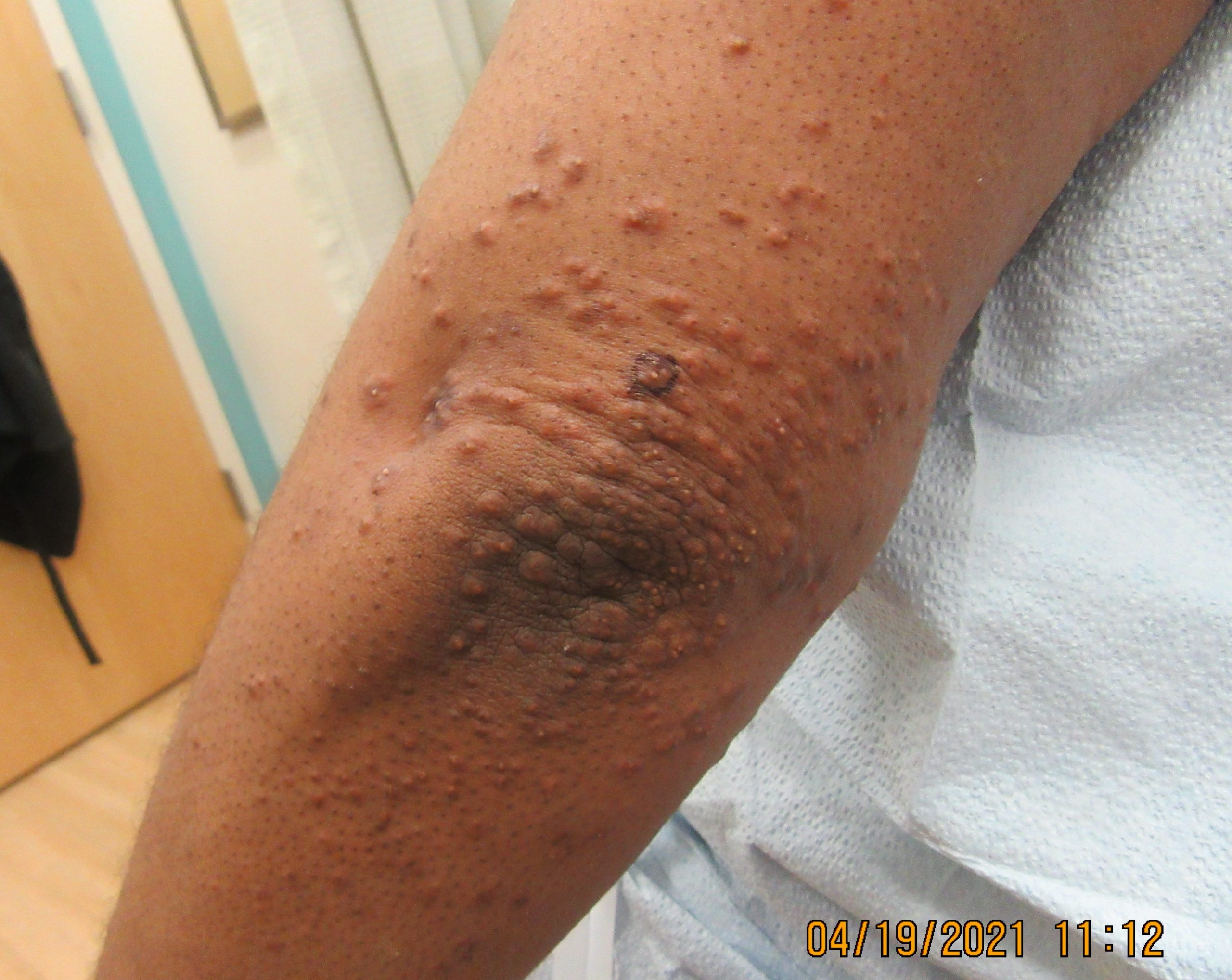
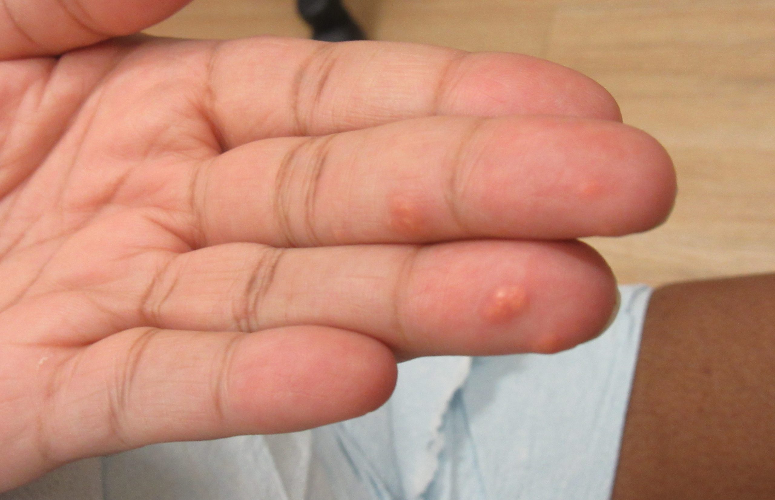
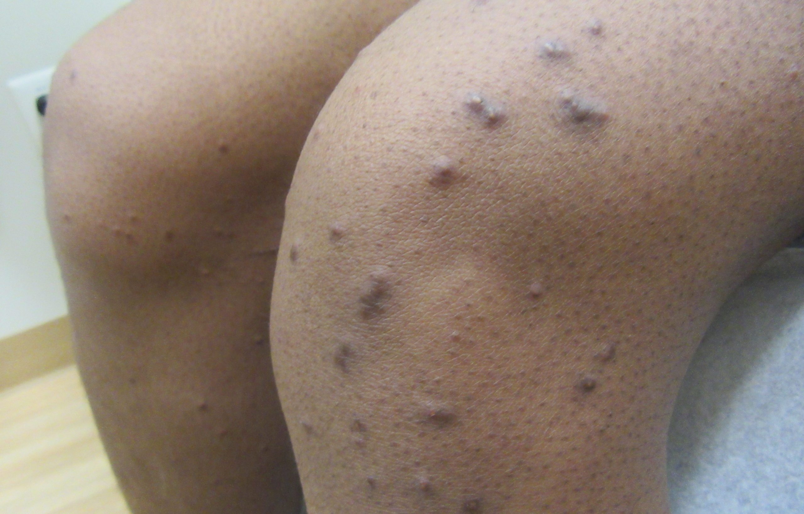
Patient is a 37 year old woman who presents with rash on the extremities for 3 months. The asymptomatic bumps started on the right elbow and subsequently spread to the left elbow and bilateral knees. She denies any other concerning symptoms.
Which of the following other laboratory findings is also most likely to be present?
A.) Elevated hemoglobin a1c
B.) Low thyroid-stimulating hormone
C.) Elevated pancreatic lipase
D.) Elevated serum sodium & osmolality along with decreased urine osmolality & specific gravity
E.) Monoclonal spike on serum protein electrophoresis
Answer: (A) Elevated hemoglobin a1c
This patient presents with crops of pink to yellow papules, some with an erythematous halo, about 1 to 5 mm in diameter distributed on the extensor surfaces of the extremities, most consistent with eruptive xanthomas.
Elevated hemoglobin a1c levels (A) above 6.5% are suggestive of diabetes mellitus, which is associated with impaired insulin activity. Deficiency or resistance to insulin both causes a decrease in lipoprotein lipase activity while thereby also contributing to hepatic overproduction of triglyceride-rich lipoproteins, both effectively serving to contribute to pathogenesis of eruptive xanthomas.1 Lipoprotein lipase is bound to capillary endothelium and is involved in both exogenous and endogenous pathways of triglyceride (TG) and cholesterol circulation. This enzyme releases free fatty acids to the peripheral tissues by catalyzing the hydrolysis of core TGs in circulating chylomicrons and VLDL molecules by way of complex interactions involving hormones, including insulin, and apoproteins, including apo C-II.1 The etiology of eruptive xanthomas is intricately related to deficiency of lipoprotein lipase activity and hepatic overproduction of TG-rich lipoproteins, both of which can result or be worsened by impaired insulin activity.1 Other environmental factors playing important roles in the appearance of eruptive xanthomas include: hypothyroidism, alcohol abuse, estrogen replacement therapy, systemic retinoids, anti-retroviral therapy, olanzapine, and azacitidine.1,2
Low thyroid-stimulating hormone (B) is suggestive of hyperthyroidism. Although hypothyroidism has been shown to be a contributing factor to the presentation of eruptive xanthomas, hyperthyroidism is not a common trigger.1,2 Common dermatologic findings associated with hyperthyroidism include pretibial myxedema, hyperhidrosis, flushing, diffuse hair thinning, onycholysis, and Plummer’s nail.3
When occurring in the context of hypertriglyceridemia, TG levels in patients with eruptive xanthomas often exceed 3,000 to 4,000.1 Acute pancreatitis, associated with elevated pancreatic lipase levels (C), occurs at a significantly increased risk in patients with very high TG levels, and has been shown to occur in approximately 10-20% in patients with TG greater than 2,000.4 Although eruptive xanthomas may be an important warning sign that may herald the onset of acute pancreatitis, the laboratory finding of elevated lipase in a patient with no other concerning symptoms would be less likely than a finding of elevated hemoglobin a1c, as diabetes mellitus is a common precipitating factor for eruptive xanthomas.
Diabetes insipidus is characterized by the laboratory findings of elevated serum sodium & osmolality along with decreased urine osmolality & specific gravity (D), and has been associated with both xanthoma disseminatum and Erdheim-Chester disease, both non-Langerhans cell histiocytoses (LCH).1 Xanthoma disseminatum is a normolipemic type of non-LCH which presents with a triad including cutaneous xanthomas, mucosal xanthomas, and diabetes insipidus.1 Xanthomas in this condition more commonly present with symmetric flexural and intertriginous involvement, however. Erdheim-Chester disease is another non-LCH which may also present with diabetes insipidus, along with fever, multiorgan involvement, other neurologic symptoms, and bone lesions and fractures.1 Although this condition may present with red to brown to yellow indurated plaques and nodules, skin involvement is infrequent in this rare condition. Both of these conditions are also much less likely in this patient with no other concerning symptoms.
Monoclonal spike on serum protein electrophoresis (E) is indicative of plasma cell dyscrasia, which has been associated with aforementioned xanthoma disseminatum, normolipemic plane xanthomas, along with number of other dermatologic conditions including Sweet’s syndrome, AL amyloidosis, necrobiotic xanthogranuloma, scleredema, scleromyxedema, IgA pemphigus, subcorneal pustular dermatosis, pyoderma gangrenosum, erythema elevatum diutinum, POEMS syndrome, Schnitzler’s syndrome, cryoglobulinemia, and Waldenstrom’s macroglobulinemia.1,5-7 Plane xanthomas classically present around the periorbital region, lateral neck, upper trunk, flexures, interdigital spaces, and palmar creases. The patient’s presentation is more consistent with eruptive xanthomas.
References
- Bolognia JL, Schaffer JV, Duncan KO, Ko CJ. Dermatology essentials E-book. Elsevier Health Sciences; 2014 Feb 26.
- Babade, M., Prodanovic, E., Mostow, E. Eruptive Xanthoma Associated with Diabetic Ketoacidosis: Lessons from a Case. Practical Dermatology. May 2006.
- Lause M, Kamboj A, Faith EF. Dermatologic manifestations of endocrine disorders. Translational pediatrics. 2017 Oct;6(4):300.
- Scherer J, Singh V, Pitchumoni CS, Yadav D. Issues in hypertriglyceridemic pancreatitis-an update. Journal of clinical gastroenterology. 2014 Mar;48(3):195.
- Iglesias-Girard L, Roy SF, Chapdelaine H, Désy D, Bouffard D, Funaro D. Disseminated Xanthosiderohistiocytosis With Monoclonal Gammopathy—A Rare Form of Xanthoma Disseminatum. JAMA dermatology. 2020 Nov 1;156(11):1270-2.
- Cohen YK, Elpern DJ. Diffuse normolipemic plane xanthoma associated with monoclonal gammopathy. Dermatology practical & conceptual. 2015 Oct;5(4):65.
- Daoud MS, Lust JA, Kyle RA, Pittelkow MR. Monoclonal gammopathies and associated skin disorders. Journal of the American Academy of Dermatology. 1999 Apr 1;40(4):507-35.