June 2021 Case Study
by Adrianna Gonzalez, MD
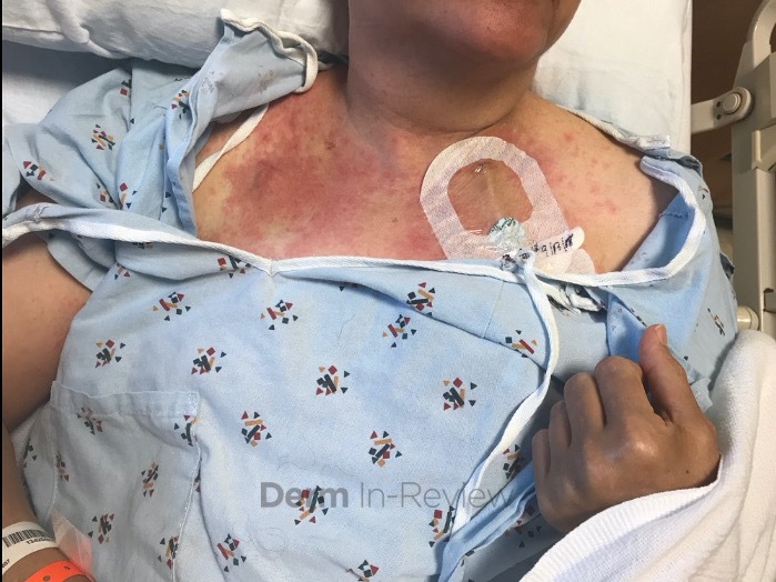
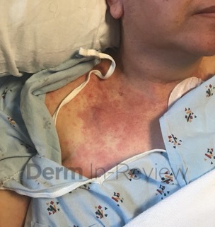
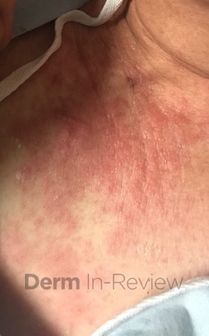
A 63-year-old woman was admitted following a hematopoietic stem cell transplant (HSCT) for the treatment of multiple myeloma. Eleven days after her transplant, she developed a morbilliform eruption on her back that subsequently spread to her chest, abdomen and upper arms (Figure 1a-b). The rash was initially pruritic but became increasingly painful. Additionally, she reported associated abdominal pain and diarrhea. Prior to HSCT, she had received high-dose melphalan, as well as induction chemotherapy with carfilzomib and dexamethasone, which had been discontinued 6 weeks prior to transplantation. Current medications included filgastrim, acyclovir and erythromycin.
Histopathological examination revealed basket weave orthokeratosis, vacuolar interface dermatitis with some necrotic keratinocytes and a sparse superficial perivascular lymphohistiocytic infiltrate with occasional melanophages and extravasated red blood cells.
Which of the following statements is FALSE regarding her condition?
A) Acral skin is often one of the first sites to be affected
B) GI and hepatic involvement is often seen with cutaneous disease
C) The most important risk factor for developing this entity is HLA compatibility
D) Due to the underlying pathophysiology, this condition is exclusively seen following allogeneic stem cell transplants as opposed to autologous stem cell transplants
E) Patients may present with erythroderma and bullae resembling SJS/TEN
Correct answer: D.) Due to the underlying pathophysiology, this condition is exclusively seen following allogeneic stem cell transplants as opposed to autologous stem cell transplants
This is FALSE. This patient has acute graft-versus-host disease, and although it is most commonly thought of as a complication of allogeneic HSCT, it may also be seen following autologous HSCT.
Explanation/Literature review:
Graft-versus-host disease is a multiorgan disorder that predominantly affects the skin, liver, GI tract (answer choice B) and lungs. Acute GVHD (aGVHD) typically presents as a morbilliform exanthem with red to violaceous lesions and an initial predilection for acral sites (answer choice A) and the upper trunk. This eruption typically develops 2-6 weeks after HSCT and may be asymptomatic or present with pruritus, pain or a burning sensation. Lesions may also present in a follicular pattern or may have a hemorrhagic appearance if the patient is thrombocytopenic. Patients with advanced stages of aGVHD (stages III-IV) may develop erythroderma and bullae, sometimes resembling toxic epidermal necrolysis (answer choice E).1 Oral manifestations such as erythema, erosions, ulcerations, lichenoid lesions or pain may occur. Chronic GVHD may present as either a lichenoid, eczematous, psoriasiform, sclerodermoid eruption and is classically defined as occurring > 100 days after HCST.
GVHD commonly occurs after the transfer of donor hematopoietic stem cells into host recipient via an allogeneic stem cell transplant. After transplantation, donor T cells recognize the immunosuppressed recipient’s tissues, resulting in the production of cytotoxic effector cells and inflammatory cytokines.2 Human leukocyte antigen compatibility between donor and recipient is therefore the most important predictor of GVHD (answer choice C). As patients undergoing autologous HSCT are not subjected to this genetic disparity, this proinflammatory response is not expected. However, GVHD has been reported to occur following autologous HSCT with an incidence between 2-13% (making answer choice D false).3-6 Notably, patients with multiple myeloma (MM) seem to be at an especially high risk of developing this syndrome.3-6 Its mechanism has been attributed to self-tolerance failure, leading to the proliferation of autoreactive CD8+ cytotoxic T cells that, with the aid of CD4+ T cells and natural killer cells, can lead to cell death.7
Management of cutaneous lesions depends on the extent of involvement and responsiveness to steroids. For limited cutaneous disease, topical corticosteroids are considered first line therapy, although topical calcineurin inhibitors may be used in some cases.8 In patients with more extensive involvement, systemic corticosteroids are considered first-line agents.9 However, corticosteroids only lead to resolution in less than 40% of patients, and disease that is refractory to steroids is associated with a significantly higher mortality risk. Extracorporeal photopheresis, mycophenolate mofetil, TNF alpha inhibitors, IL-2 receptor antibodies, antithymocyte globulin and phototherapy have been used with varying success.9 Recently, studies have reported successful management of GVHD with Janus kinase (JAK) inhibitors such as ruxolitinib, although more studies are needed to elucidate their efficacy and safety for this purpose.10
References
- Hausermann P, Walter RB, Halter J, et al. Cutaneous graft-versus-host disease: a guide for the dermatologist. Dermatology. 2008;216(4):287-304.
- Schmaltz C, Alpdogan O, Muriglan SJ, et al. Donor T cell-derived TNF is required for graft-versus-host disease and graft-versus-tumor activity after bone marrow transplantation. Blood. 2003;101(6):2440-2445.
- Lazarus HM, Sommers SR, Arfons LM, et al. Spontaneous autologous graft-versus-host disease in plasma cell myeloma autograft recipients: flow cytometric analysis of hematopoietic progenitor cell grafts. Biol Blood Marrow Transplant. 2011;17(7):970-978.
- Fidler C, Klumpp T, Mangan K, et al. Spontaneous graft versus host disease occurring in a patient with multiple myeloma after autologous stem cell transplant. Am J Hematol. 2012;87(2):219-221.
- Alonso S, Cabrero M, Caballero JC, et al. Acute graft-versus-host disease and bronchiolitis obliterans after autologous stem cell transplantation in a patient with multiple myeloma. Clin Case Rep. 2015;3(6):370-375.
- Lee SE, Yoon JH, Shin SH, Park G, Min CK. Skin Graft-versus-host Disease Following Autologous Stem Cell Transplantation for Multiple Myeloma. Immune Netw. 2013;13(3):107-110.
- Miura Y, Thoburn CJ, Bright EC, Hess AD. Cytolytic effector mechanisms and gene expression in autologous graft-versus-host disease: distinct roles of perforin and Fas ligand. Biol Blood Marrow Transplant. 2004;10(3):156-170.
- Penas PF, Fernandez-Herrera J, Garcia-Diez A. Dermatologic treatment of cutaneous graft versus host disease. Am J Clin Dermatol. 2004;5(6):403-416.
- Strong Rodrigues K, Oliveira-Ribeiro C, de Abreu Fiuza Gomes S, Knobler R. Cutaneous Graft-Versus-Host Disease: Diagnosis and Treatment. Am J Clin Dermatol. 2018;19(1):33-50.
- Zeiser R, Burchert A, Lengerke C, et al. Ruxolitinib in corticosteroid-refractory graft-versus-host disease after allogeneic stem cell transplantation: a multicenter survey. Leukemia. 2015;29(10):2062-2068.