
August 2021 Case Study
by Jessica Kalen, MD
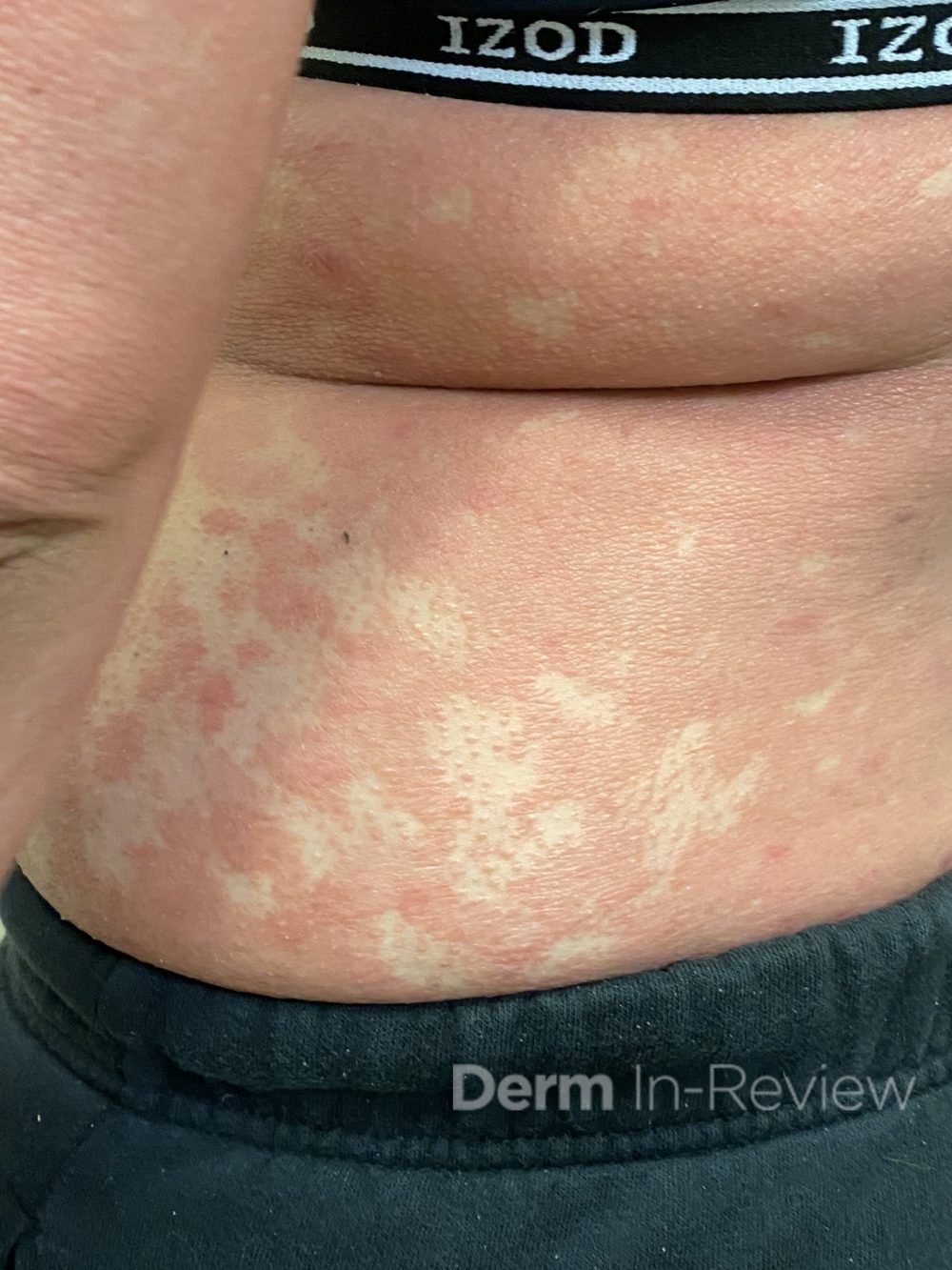
A 37-year-old female with no past medical history presents for evaluation of the pictured rash over the body. Initially began 6 weeks ago with significant associated pruritus. Prior to presentation was treated with methylprednisolone taper and triamcinolone 0.1% ointment. Additional clinical findings include a keratoderma of the bilateral hands and yellowing of fingernails with subungual debris. She denies joint pain or stiffness, but does report malaise and chills. She has no personal or family history of skin conditions.
Based on the clinical presentation, what are the anticipated histopathological findings for this condition?
A.) Regular acanthosis with neutrophilic debris and parakeratosis in stratum corneum
B.) Shoulder parakeratosis and neutrophilic debris focused around follicular ostia
C.) Alternating vertical and horizontal orthokeratosis and parakeratosis, acanthosis, and mild spongiosis
D.) Subacute spongiosis with extravasation and transepidermal elimination of red blood cells
E.) Lichenoid interface dermatitis with hypergranulosis and dyskeratosis
Correct answer: C
Explanation
Based on the clinical presentation, the anticipated histopathological findings would be most consistent with pityriasis rubra pilaris (PRP). This condition presents with widespread waxy, orange plaques and islands of sparing as well as prominent follicular hyperkeratosis, often compared to a nutmeg grater.1 Onset occurs over the course of several weeks with an, often, rapid progression to erythroderma.1 PRP is classified into six subtypes based on age, duration, and clinical appearance. Subtypes include classic adult, atypical adult, classic juvenile, circumscribed juvenile, atypical juveline, and HIV-associated.1
Histopathologically, the classic finding of PRP is alternating orthokeratosis and parakeratosis both vertically and linearly.2 This is often compared to a “checkboard”. Additionally, there is follicular plugging with shoulder parakeratosis, hypergranulosis, mild spongiosis, and sparse lymphohistiocyctic infiltrate.1
Standardized treatment guidelines of PRP remain to be established. However, several systemic medications have been empirically utilized to treat this condition. PRP has been successfully treated with oral retinoids, methotrexate, cyclosporine, and azathioprine.1 Now with the advent of biologics, PRP has also shown good response to TNF-alpha inhibitors, guselkumab, ixekizumab, secukinumab.1,3,4
The remaining answers do not describe the histopathological findings of PRP. Choice A describes findings of psoriasis, choice B describes seborrheic dermatitis, choice D describes pityriasis rosea, and choice E describes lichen planus.
References
- Bolognia JL, Schaffer JV, Cerroni L. Dermatology. 4th Philadelphia. Elsevier Limited.
- Elston DM, et al. Dermatopathology. 4th 2019. Elsevier.
- Pilz AC, et al. Treatment of pityriasis rubra pilaris with guselkumab. JAMA Dermatol. Dec 2019;155(12):1424-1426.
- Haynes D, et al. Evaluation of ixekizumab treatment for patients with pityriasis rubra pilaris: a single-arm trial. JAMA Dermatol. 2020;156(6):668-675.