April 2021 Case Study
by Gabrielle Schwartzman, MD
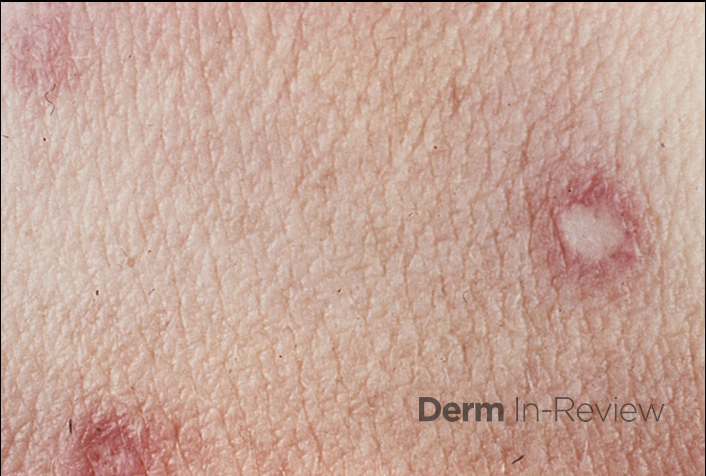
A 32-year-old female with past medical history of systemic lupus erythematous on prednisone, presents with an 8 month history of “painful, cracking, red skin lesions with a white center.” Patient was referred by her rheumatologist for evaluation of these skin lesions of the bilateral dorsum of hands and knees. Patient had since been using topical steroid ointment without improvement. The lesions are painful. Physical exam showed multiple erythematous papules coalescing into plaques and many white atrophic scar-like papules within these plaques on the bilateral hands and knees. Dermoscopy of the lesions showed porcelain white atrophic centers with a rim of telangiectasias.
Based upon the patient history and clinical examination, the lesions are most likely associated with of the following processes?
A.) Thrombo-obliterative vasculopathy
B.) Autoantibodies specific for hemidesmosomal antigens
C.) Primary viral infection
D.) Genetic mutation of TGM1
E.) Autoimmunity targeting melanocyte destruction
Correct answer: A) Thrombo-obliterative vasculopathy
Degos disease, also referred to as malignant atrophic papulosis, is a rare small vessel arteriopathy with a pathognomonic appearance of atrophic porcelain-white central papules surrounded by telangiectasias.1 Degos disease can be idiopathic or secondary to autoimmune disorders or connective tissue diseases including antiphospholipid antibody syndrome, systemic sclerosis, dermatomyositis, and SLE, or viral infection.1,2
Proposed pathology of Degos disease includes coagulopathy, endothelial cell damage, and vasculitis, however the exact mechanism of not well established.3,4 The effectiveness of eculizumab, a terminal complement inhibitor, suggests a complement-mediated process. Some have suggested that Degos-like lesions may be a common end point to a variety of vascular insults.5 The histology is not consistent but often shows a wedge-shaped connective tissue necrosis in the deep corium due to a thrombotic occlusion of the small arteries.6 Systemic disease mostly occur at the intestine and central nervous system.6
Diagnosis is based on the characteristic skin lesions, papular skin lesions with central porcelain-white atrophy and surrounding teleangiectatic rim.6 Less than 200 cases have been reported. The first manifestation usually occurs between the 20th and 50th year of life.6 Cases of Degos-like lesions associated with SLE have been reported in the literature.3 The disease course, management, and prognosis of these cases have varied.
References
- Chieosilapatham P, Prinyaroj N, Jamjanya S, et al. Degos-like lesions as a cutaneous manifestation of cytomegalovirus infection: A rare and serious complication in a patient with drug-induced hypersensitivity syndrome. J Dermatol. 2020;(August):1-4. doi:10.1111/1346-8138.15717
- Vinay K, Sawatkar G, Dogra S, Saikia UN. Systemic lupus erythematosus with Degos disease: role of dermatoscopy in diagnosis. Int J Dermatol. 2017;56(7):770-772. doi:10.1111/ijd.13629