Krista Reznik – Page 5 – Derm In-Review

September 2022 Case Study

By 2022 Case Studies

September 2022 Case Study

by Alexis Carrington, MD


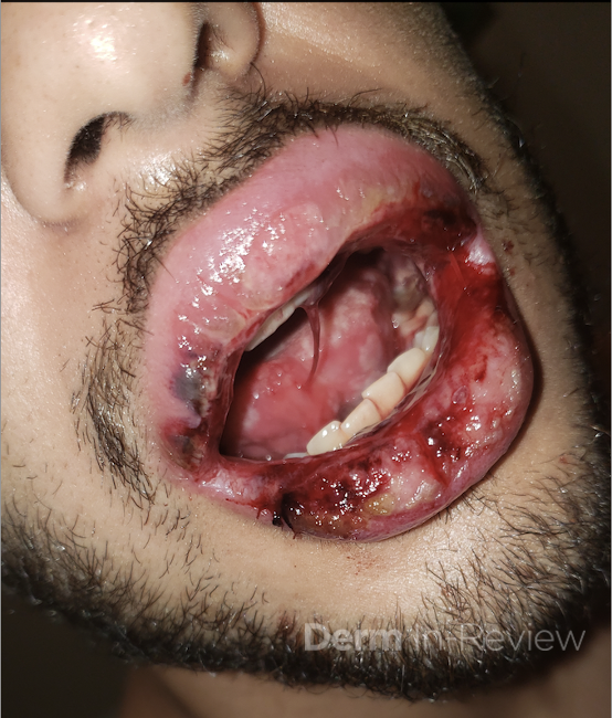
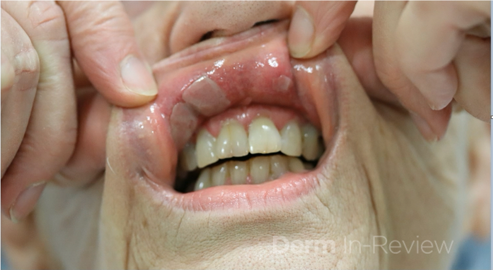
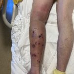
A 22 year-old male with a past medical history of asthma presents to the ED with a rash on the mouth and bilateral palms that appeared a day prior to presentation. A week prior, he went to the ED for worsening throat and mouth pain, where he was diagnosed with thrush and given nystatin but didn’t have any skin manifestations at that time. He also reported subjective fevers, chills, decreased appetite, fatigue and non-bloody diarrhea that started two days ago. Exam showed hemorrhagic mucositis with some minor re-epithelialization and a papular target lesions on the hands and left arm. Based on examination, a diagnosis of Erythema Multiforme was made.

What is the most common precipitating factor for Erythema Multiforme?

A.) Herpes Simplex Virus (HSV)
B.) Parapoxvirus
C.) Mycoplasma pneumoniae
D.) Histoplasma capsulatum


Correct answer: A.) Herpes Simplex Virus (HSV)

Erythema Multiforme (EM) is an immune-mediated mucocutaneous condition characterized by “target” lesions. Severe mucosal involvement is what distinguishes erythema multiforme major from erythema multiforme minor. EM episodes can be recurrent or persistent, but are typically self-limiting. Some triggers of EM include infections, as well as medications and even vaccinations.

The characteristic morphology of EM are typical target lesions, which are a round shape with a well defined border and consist of three distinct zones distributed on the extremities and the face, the dorsal hands and forearms being the most frequently involved. Mucosal lesions of EM are vesiculobullous and develop into painful erosions on the buccal mucosa and lips. Diagnosis is clinical but not based solely on histological findings.

The most common associated infectious agent with EM is Herpes Simplex Virus (answer A). Parapoxvirus (answer B) Histoplasma capsulatum (answer D) and Mycoplasma pneumoniae (answer C) are observed less frequently.

Treatment is supportive as EM episodes are typically self limiting. However, early therapy with systemic corticosteroids can be considered in severe forms of EM with functional impairment.


References

  1. Canavan TN, Mathes EF, Frieden I, Shinkai K. Mycoplasma pneumoniae-induced rash and mucositis as a syndrome distinct from Stevens-Johnson syndrome and erythema multiforme: a systematic review. J Am Acad Dermatol. 2015;72(2):239–45. doi:10.1016/j.jaad.2014.06.026.
  2. Soares A, Sokumbi O. Recent Updates in the Treatment of Erythema Multiforme. Medicina (Kaunas). 2021;57(9):921. Published 2021 Sep 1. doi:10.3390/medicina57090921.
  3. Bolognia, Jean L. Dermatology volume 2. Mosby, 2018.

August 2022 Case Study

By 2022 Case Studies

August 2022 Case Study

by Blair Allais, MD

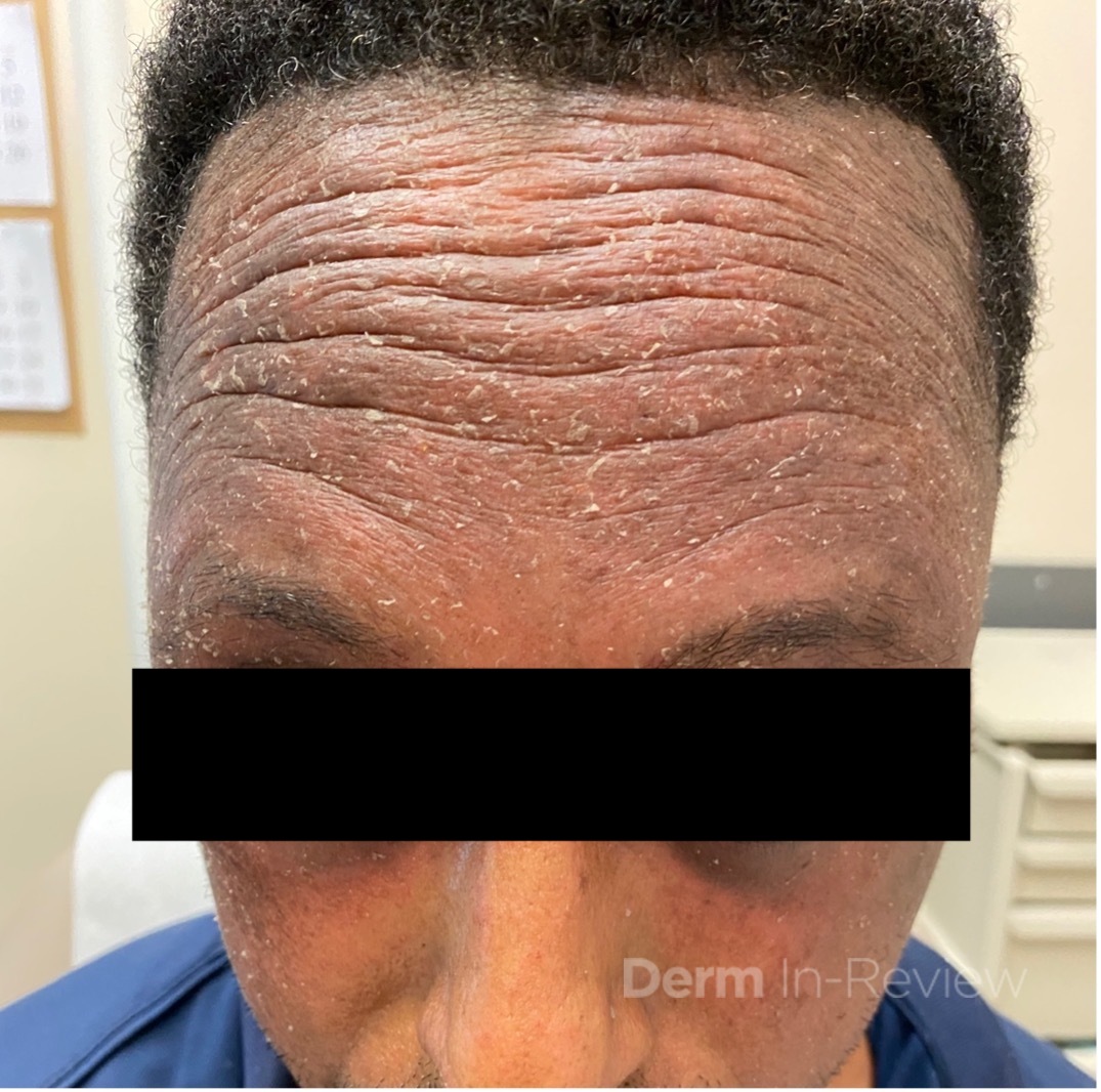
A 55-year-old male presented to clinic with a new worsening red, scaly rash of the forehead, temples and upper cheeks. He had tried to use over the counter hydrocortisone and witch hazel toner at home without improvement of the rash. On further questioning of possible exposures, the patient endorsed switching his home hair dye recently and used it for the second time several days prior to the development of the rash.

Which of the following cross reacts with the likely culprit allergen in this case?

A.) Aspirin
B.) Penicillin
C.) Glipizide
D.) Lidocaine
E.) Furosemide

Correct answer: C.) Glipizide

Para-phenylenediamine (PPD) dye is commonly found in many consumer products including hair dye, black henna tattoos, textiles, leather, fur, printer ink, and black rubber products.1 The majority of reactions to topical PPD are delayed-type IV hypersensitivity in nature and characterized by pruritus, erythema, and vesiculobullous eruptions that may occur on the face, scalp margins, or ears.2 Cross-sensitization to other compounds that also contain a benzene ring at the para position can occur.3 A helpful mnemonic to remember the chemicals that cross-react with PPD can be found in Chapter 2 of the Derm-In-Review Text: “AA SSTEPP”, which stands for Azo, Aniline dyes, Sulfonamides, Sulfonylurea, Thiazides, Esters (benzocaine), Procainamide, PABA and PABA esters.


Glipizide is a sulfonylurea and cross reacts with PPD. The other answer choices are distractors and incorrect. Para-aminosalicylic acid (an anti-tuberculosis medication) is a cross reactor to PPD dye, and one should not confuse para-aminosalicylic acid with aspirin (acetyl salicylic acid). While sulfonamides cross react with PPD, other antibiotics such as penicillin do not. While ester anesthetics (procaine, chlorprocaine and tetracaine) cross react with PPD, amide antibiotics such as lidocaine do not.  While thiazide diuretics (hydrochlorothiazide, chlorthalidone) cross react with PPD, loop diuretics such as furosemide do not.


References

  1. Rietschel RL, Fowler JF. Fishers Contact Dermatitis, 4th edn. Baltimore, MD: Williams and Wilkins, 1995.
  2. Mukkanna KS, Stone NM, Ingram JR. Para-phenylenediamine allergy: current perspectives on diagnosis and management. J Asthma Allergy. 2017;10:9-15. Published 2017 Jan 18. doi:10.2147/JAA.S90265
  3. McFadden JP, Yeo L, White JL. Clinical and experimental aspects of allergic contact dermatitis to para-phenylenediamine. Clin Dermatol. 2011;29(3):316-324. doi:10.1016/j.clindermatol.2010.11.011

July 2022 Case Study

By 2022 Case Studies

July 2022 Case Study


A 70-year-old woman with history of hypothyroidism and asthma presents to clinic with a 3-month history of sores in her mouth. She denies any other symptoms, but she does report a 5-pound weight loss due to pain with eating. On further exam, there are erosions of the gingiva, buccal mucosa, and posterior neck. There is no involvement of other mucosal sites. Patient undergoes biopsies for hematoxylin and eosin staining and direct immunofluorescence (DIF). Results showed a subepidermal bullae with positive IgG and C3 staining at the dermo-epidermal junction. Further testing revealed presence of anti-laminin 332 antibodies.

What additional work up is needed?

A.) Referral to ophthalmology
B.) Pulmonary function testing
C.) Perform all age-appropriate cancer screenings
D.) Endoscopy
E.) No additional work up needed

Correct answer: C.) Perform all age-appropriate cancer screenings

Mucous membrane pemphigoid (MMP) is a subepidermal autoimmune blistering disorder with various phenotypes. As the name suggests, clinical features involve erosions of mucosal surfaces, often with subsequent scarring. The most commonly affected mucosal sites are oral and ocular surfaces. Those with ocular involvement are at risk for blindness so it is important to ensure ophthalmologic monitoring when necessary. Non-mucosal skin can be affected, but this occurs in less than 50% of patients.1


Previously, subtypes were classified based on antigenic targets or area of involvement – anti-elipigrin cicatricial MMP (laminin 332), ocular MMP (b4 subunit of integrin a6b4), anti-bullous pemphigoid antigen MMP (BPAG180 or BPAG230).1 Additionally, the “Brunsting-Perry” variant classified a subset of patients with cutaneous involvement of the head and neck resulting in a cicatricial alopecia.1 However, recent updates in nomenclature have moved away from use of these subtypes and toward use of phenotypic descriptions.2


Diagnosis of MMP requires a combined approach as diagnosis can often be delayed. As with other vesiculobullous diseases, DIF is essential to diagnosis and has been found to be the most sensitive diagnostic tool.3,4 Classic findings on DIF are IgG, IgA, and/or C3 deposition at the dermo-epidermal junction.4 Salt split skin technique is useful to differentiate likely antigenic targets. Roof staining with IgG or IgA is consistent with BP180 or BP230 targets while floor staining with IgG or IgA correlates to laminin 332 or type VII collagen targets.4 It is important to remember that anti-laminin 332 MMP, as seen in this patient, is associated with an increased risk of malignancy.1,4  Thus, it is important that patients with this variant undergo age-appropriate cancer screenings at the time of diagnosis.


References

  1. Bolognia JK, Schaffer JV, Cerroni L. Dermatology. Philadelphia: Elsevier. 2018.
  2. Rashid H, et al. European guidelines (S3) on diagnosis and management of mucous membrane pemphigoid, initiated by the European Acadmy of Dermatology and Venereology – Part I. 2021;35:1750-1764.
  3. Rashid H et al. Assessment of diagnostic strategy of mucous membrane pemphigoid. JAMA Derm. July 2021;157(7):780-787.
  4. Schmidt E, et al. European guidelines (S3) on diagnosis and management of mucous membrane pemphigoid, initiated by the European Academy of Dermatology and Venereology – Part II. JEADV. 2021;35:1926-1948.

June 2022 Case Study

By 2022 Case Studies

June 2022 Case Study

by Adrianna Gonzalez, MD

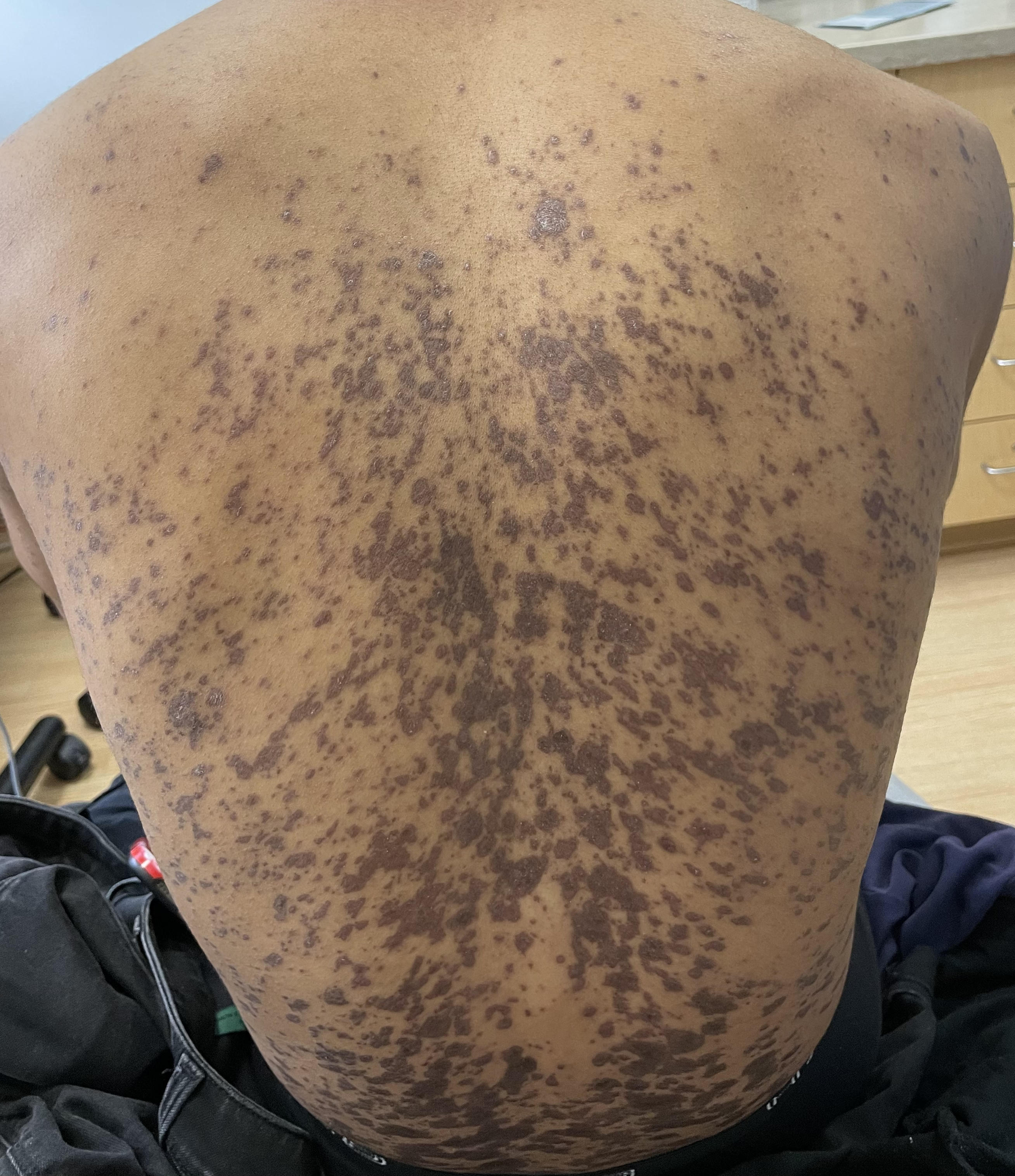
A 29 year old male with no significant past medical history presented with complaints of a generalized rash which began two months prior. He stated the eruption began as a few lesions in the trunk that progressively grew in number and generalized to involve upper and lower extremities, buttocks, and genitals. He stated rash was associated with significant itch. Of note, he stated he had taken a new medication several weeks prior to the onset of his rash. Findings on clinical examination can be seen in Figure 1. A 4mm punch biopsy of a lesion on his mid back was performed. Histopathological examination revealed orthokeratosis, hypergranulosis, acanthosis with sawtoothing rete ridge pattern, dyskeratotic keratinocytes and a band-like lymphocytic infiltrate.

Which of the following statements is FALSE regarding this patient’s condition?

A.) The oral mucosa is the most commonly involved body site in this entity
B.) On histology, dyskeratotic keratinocytes are classically found at the upper level of the epidermis
C.) Direct immunofluorescence may be positive for “shaggy” fibrinogen deposits along the dermoepidermal junction
D.) Flares may be triggered by certain contact allergens including copper
E.) The majority of cases will remit within 2 years

Correct answer: B.) On histology, dyskeratotic keratinocytes are classically found at the upper level of the epidermis

The clinical and histopathological findings in this case are consistent with lichen planus (LP). On histology, LP is characterized by compact orthokeratosis, wedge-shaped hypergranulosis, sawtooth pattern of rete ridges, vacuolar degeneration of the basal layer, dyskeratotic keratinocytes (Civatte bodies), and a band-like lymphocytic infiltrate at the dermoepidermal junction (DEJ). Dyskeratotic keratinocytes are classically present at the lower levels of the epidermis and at the superficial papillary dermis near the basal layer, the site of destruction.1 Dyskeratotic keratinocytes can be seen in the suprabasilar, upper epidermal layer in erythema multiforme, Stevens-Johnson Syndrome and toxic epidermal necrolysis.2 Direct immunofluorescence (DIF) demonstrates “shaggy” deposition of fibrinogen along the basement membrane zone, as well as staining of colloid bodies with one or more of IgM, IgG, IgA and C3  (answer C).


Lichen planus is an inflammatory disorder of the skin, mucosa, hair and nails. Although the incidence appears to vary throughout geographical areas, the oral mucosa has been found to be the most commonly involved body site in most study populations (answer A).1,3 Oral lesions can be observed in over 50% of patients with LP, and can often be the only site of involvement.2 A recent epidemiological study estimated that the global pooled prevalence of oral LP was 0.89% among the general population and 0.98% among clinical patients.4 Cutaneous LP is thought to affect < 1% of the population worldwide and commonly presents as pruritic, purple, polygonal, flat-topped papules commonly involving the extremities and lower back.5 There are numerous variants of cutaneous LP, including actinic, annular, atrophic, bullous, drug-induced, genital, hypertrophic, inverse, linear, LP pigmentosus and lichen planopilaris. LP may also involve the nails and present with longitudinal ridging, trachyonychia, thinning, dorsal pterygium and even 20 nail dystrophy. While its pathophysiology has not been fully elucidated, LP is thought to be caused by an altered cellular immune response, in which cytotoxic CD8+ T cells attack basal keratinocytes. LP has been associated with various triggers, including infections such as hepatitis C virus (more common in oral erosive LP), medications, contact allergens, vaccinations (hepatitis B virus and influenza) and genetics. Implicated contact allergens include gold, mercury amalgam and copper (answer D).1  Most cases of LP self-resolve within 1-2 years (answer E). Oral LP (especially oral erosive LP), nail LP and hypertrophic LP tend to be persistent and recalcitrant to treatment.

References

  1. Boch K, Langan EA, Kridin K, Zillikens D, Ludwig RJ, Bieber K. Lichen Planus. Front Med (Lausanne). 2021;8:737813.
  2. Bolognia J, Jorizzo, J. L., & Schaffer, J. Dermatology. Fourth Edition ed. Philadelphia: Elsevier Saunders; 2012.
  3. Arnold DL, Krishnamurthy K. Lichen Planus. In: StatPearls. Treasure Island (FL)2022.
  4. Li C, Tang X, Zheng X, et al. Global Prevalence and Incidence Estimates of Oral Lichen Planus: A Systematic Review and Meta-analysis. JAMA Dermatol. 2020;156(2):172-181.
  5. Tziotzios C, Lee JYW, Brier T, et al. Lichen planus and lichenoid dermatoses: Clinical overview and molecular basis. J Am Acad Dermatol. 2018;79(5):789-804.

May 2022 Case Study

By 2022 Case Studies

May 2022 Case Study

by Emily Murphy, MD

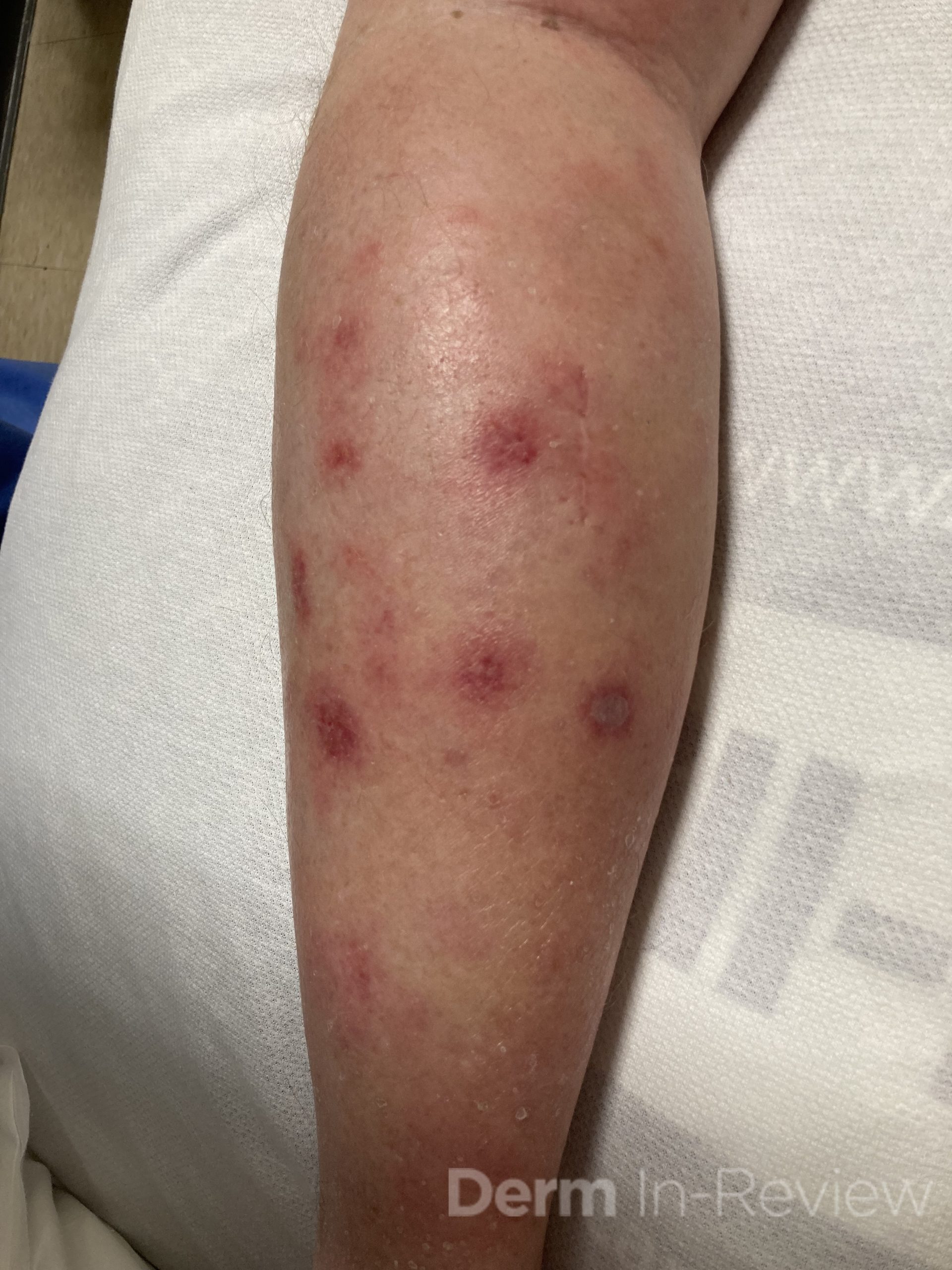
A 65-year-old male with a past medical history of HIV (CD4 count 250, undetectable viral load) and chronic congestion presented with a chief complaint of recurrent cellulitis of the left lower leg. The redness and swelling persisted for 3 months despite antibiotics. On presentation, he had circumferential edema and induration of the left lower leg with erythematous, tender nodules and early bullae (figure 1). A week later, his disease progressed with ulceration and necrosis of these tender nodules (figure 2). A punch biopsy was done, which showed an atypical lymphoid infiltrate in the dermis and subcutaneous tissue with an angiocentric pattern, composed of large, pleomorphic cells. Extensive necrosis was present with karyorrhetic debris. Immunostains were positive for T cells (CD2), natural killer cells (CD56), and cytotoxicity (granzyme B). A biopsy of the nasal septum showed similar findings. The patient was diagnosed with extranodal NK/T cell lymphoma, nasal type.

Which virus is part of the diagnostic criteria for this lymphoma?

A.) Human herpesvirus-8
B.) Polyomavirus
C.) Human immunodeficiency virus
D.) Ebstein Bar Virus
E.) Cytomegalovirus

Correct answer: D.) Ebstein Bar Viru

Extranodal NK/T cell lymphoma, nasal type is an aggressive, non-Hodgkin lymphoma that has four criteria according to the World Health Organization: vascular damage, prominent necrosis, a cytotoxic phenotype, and association with the Ebstein bar virus (EBV, answer D).1,2 EBV positivity is seen in the both the skin and serum. Pathologically, the atypical lymphocytes are positive for cytotoxic markers (granzyme B, peforin), natural killer cell markers (CD2), T cell markers (cytoplasmic CD3), and EBV.1 Extranodal NK/T cell lymphoma is rare in the United States, but its incidence is increasing. It is more common in East Asia and Latin America.1

This lymphoma typically affects the upper aerodigestive tract, causing congestion, epistaxis, or necrotizing lesions of the nose or hard palate.1 Other sites can be involved, including the extranasal skin, gastrointestinal tract, testes, or lungs.1,3,4 On the skin, either a generalized morbilliform eruption or purpuric nodules with or without ulceration can be seen.3 Hemophagocytic lymphohistiocytosis can occur, but is rare, occurring in only 3% of patients.1

NK/T cell lymphoma has an increased incidence in patients with HIV; a study of 93 patients with this lymphoma found that it occurred at a 15 times higher incidence in patients with HIV compared to the general population.5 However, HIV (choice C) is not part of the diagnostic criteria like EBV. Human herpesvirus-8 (Kaposi sarcoma), Polyomavirus (Merkel Cell Carcinoma), and cytomegalovirus are not associated with Extranodal NK/T cell lymphoma (choices A, B, E).

Treatment is determined by the Ann Arbor Staging and various prognostic tools, like the PINK-E system (age > 60 years, Stage III/IV, nonnasal primary localization, distant lymph node involvement, and detectable plasma EBV DNA).6 Depending on the stage and prognosis, treatment is typically with an asparaginase-based regimen called SMILE (dexamethasone, methotrexate, ifosfamide, L-asparaginase, and etoposide).1,6,7 NK/T cell lymphoma cells are unable to synthesize the amino acid, asparagine, and as this amino acid is depleted with asparaginase, protein synthesis is disrupted and the cells ultimately undergo apoptosis.6 Chemotherapy is typically followed by involved field radiation therapy.1  Various immunotherapies are currently being studied for patients with recalcitrant or relapsed disease as well, including programmed death 1 inhibitors, JAK 3 inhibitors, or anti-CD30 therapies.8 Response to therapy can be monitored with EBV DNA titers, which should decrease with treatment.1

References

  1. Haverkos BM, Pan Z, Gru AA, et al. Extranodal NK/T Cell Lymphoma, Nasal Type (ENKTL-NT): An Update on Epidemiology, Clinical Presentation, and Natural History in North American and European Cases. Curr Hematol Malig Rep. 2016;11(6):514-527. doi:10.1007/s11899-016-0355-9
  2. Willemze R, Cerroni L, Kempf W, et al. The 2018 update of the WHO-EORTC classification for primary cutaneous lymphomas. Blood. 2019;133(16):1703-1714. doi:10.1182/blood-2018-11-881268
  3. Orlowski GM, Tan AJ, Evan-Browning E, Scharf MJ. Primary cutaneous nasal-type NK/T-cell lymphoma presenting as purpuric nodules on the lower leg. JAAD Case Rep. 2020;6(10):1075-1078. doi:10.1016/j.jdcr.2020.08.007
  4. Rahal A, Reddy PS, Alvares C. Extranodal NK/T-Cell Lymphoma, Nasal Type, Presenting as a Breast Mass. Cureus. 7(12):e408. doi:10.7759/cureus.408
  5. Castillo J, Pantanowitz L. HIV-Associated NK/T-Cell Lymphomas: A Review of 93 Cases. Blood. 2007;110(11):3457-3457. doi:10.1182/blood.V110.11.3457.3457
  6. van Doesum JA, Niezink AGH, Huls GA, Beijert M, Diepstra A, van Meerten T. Extranodal Natural Killer/T-cell Lymphoma, Nasal Type: Diagnosis and Treatment. Hemasphere. 2021;5(2):e523. doi:10.1097/HS9.0000000000000523
  7. Yamaguchi M, Kwong YL, Kim WS, et al. Phase II study of SMILE chemotherapy for newly diagnosed stage IV, relapsed, or refractory extranodal natural killer (NK)/T-cell lymphoma, nasal type: the NK-Cell Tumor Study Group study. J Clin Oncol. 2011;29(33):4410-4416. doi:10.1200/JCO.2011.35.6287
  8. Lv K, Li X, Yu H, Chen X, Zhang M, Wu X. Selection of new immunotherapy targets for NK/T cell lymphoma. Am J Transl Res. 2020;12(11):7034-7047.

April 2022 Case Study

By 2022 Case Studies

April 2022 Case Study

by Blair Allais, MD

A 63-year-old male presented to clinic with a 2-year history of asymptomatic annular hypopigmented plaques on the trunk and extremities that eventually became erythematous to violaceous. He had been prescribed topical steroids by an outside practitioner without improvement in the lesions. Biopsy performed on one of the plaques revealed a granulomatous reaction surrounding adnexal structures and in the papillary dermis. Fite stain revealed numerous pink to red acid-fast organisms within the granulomas ultimately rendering a diagnosis of lepromatous leprosy. The patient was started on therapy with Rifampin 600mg monthly, Minocycline 100mg monthly and Moxifloxacin 400mg monthly. Approximately 3 months after initiating treatment the patient developed new nodular skin lesions on the extensor arms and medial thighs associated with fever, myalgias, fatigue and joint pain.

What is the best course of treatment for this patient’s new presentation?

A.) Prednisone
B.) Clofazimine
C.) Thalidomide
D.) Methotrexate

Correct answer: C) Thalidomide

The patient described has a diagnosis of lepromatous leprosy and is experiencing a reactional state to treatment. Approximately 50% of patients with leprosy will experience a reaction after the institution of therapy. Aside from initiating antibiotic therapy, other causes of reactional states include intercurrent infections, vaccination, pregnancy, vitamin A, iodides and bromide. Reactions are an important cause of permanent nerve damage in borderline patients and are abrupt in appearance.


Two major types of reactional states have been described. Type 1 reactions due to enhancement of cell-mediated immunity with a Th1 cytokine pattern (IFN-y, TNF, IL-1ß, IL-2, IL-12). These reactions are considered to reversal reactions with “upgrading” and are seen predominantly in borderline or tuberculoid forms of leprosy. They are characterized by increased inflammation within existing skin lesions in addition to acute nerve pain or tenderness (neuritis) and loss of function. Treatment of choice for these reactions is prednisone (A) starting at a dose of 40 – 60 mg/day. Neuritis and eye lesions are urgent indications for therapy and dose and duration is determined by the clinical course of the reaction. Steroids are tapered slowly over months to years once the reaction is controlled. In some cases, clofazimine (B) may be added as it has some activity against the type 1 reaction, and may be added in doses of up to 300 mg/day as tolerated.


Type 2 reactions are due to excessive humoral immunity with a Th2 cytokine pattern (IL-4, IL-10) and characterized by formation of immune complexes. They are seen predominately in patients with lepromatous and borderline lepromatous forms of leprosy, especially in patients with a high bacterial load who are undergoing treatment. Multisystem involvement is seen with systemic symptoms including nodular skin lesions, fever, myalgias, malaise, severe joint swelling and pain, iridocyclitis, lymphadenitis, hepatosplenomegaly, orchitis and glomerulonephritis. The intensity of the reaction can vary from mild to severe and can last from a few days to weeks, months, or even years. Pathology is characterized by cutaneous and systemic small vessel vasculitis, and treatment of choice is Thalidomide (C). According to the newly developed U.S. regimen for treatment for lepromatous leprosy, methotrexate (D) 15mg weekly can be prescribed to prevent reactions at the time of initiating multi drug treatment.

References

  1. Ramos-e-Silva M, Ribeiro de Castro MC. Chapter 75: Mycobacterial Infections. 4th Ed. Philadelphia, PA: Elsevier; 2018.
  2. James WD et al. Chapter 17: Hansen Disease. Andrews’ Diseases of the Skin. 13th Philadelphia, PA: Elsevier; 2020.
  3. Bennett JE et al. Principles and Practice of Infectious Diseases. 6th edit.P.2887-2896

March 2022 Case Study

By 2022 Case Studies

March 2022 Case Study

by Daniel Nussbaum, MD

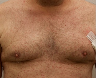
A 50-year-old man with no past medical history presents with the pictured rash on his chest, arms, abdomen, and back for five years. Although asymptomatic, the patient reported increasing redness under hot water without associated pruritus. He denied any systemic symptoms. Examination showed monomorphic erythematous macules and papules distributed diffusely over the chest, upper extremities, abdomen, and back. He denies any systemic symptoms and has no personal or family history of skin conditions other than the pictured lesions.

Which of the following laboratory tests would aid in confirming the diagnosis in this patient?

A.) Indirect immunofluorescence
B.) Anti-gliadin Ab
C.) Anti-nuclear Ab
D.) Anti-Jo-1 Ab
E.) Serum tryptase

Correct answer: E

The history and image provided are most consistent with a adult onset cutaneous mastocytosis. Histopathology of cutaneous mastocytosis commonly reveals mast cells concentrated in the dermal papillae, which can be confirmed with staining for mast cells (see below).1 The patient was recommended to avoid triggers for mast cell degranulation such as heat, sunlight, stress, medications, and alcohol. Additionally, he was referred to hematology for bone marrow biopsy, which was deferred due to lack of systemic symptoms.

Mastocytosis is a spectrum of diseases with various cutaneous and systemic manifestations. Cutaneous findings include flushing, pruritus, urticaria, and bullae. Systemic symptoms include bone pain, headaches, fatigue, nausea, vomitus, diarrhea, weight loss, cramping, chest pain, palpitations, and syncope.1 Patients with mastocytosis, especially the systemic variant, frequently have a mutation in KIT D816V, a tyrosine kinase receptor. Tissue diagnoses of mastocytosis can be confirmed with Giemsa, CD117, Leder, toluidine blue, and tryptase stains.

Diffuse cutaneous mastocytosis is a rare subset of mastocytosis, usually presenting in childhood low risk of systemic involvement. However, when presentation occurs in adulthood, systemic involvement should be evaluated with serum tryptase and bone marrow biopsy when indicated. Without a bone marrow biopsy, the risk of systemic involvement can be estimated from symptoms and a tryptase level.2 Based on clinical presentation and laboratory findings, physicians must consider working up systemic involvement to avoid morbidity and mortality.3,4

Indirect immunofluorescence (IIF) (answer A) is useful in the diagnosis of blistering dermatoses. Specifically, IIF of serum Ab to monkey esophagus can be useful in the diagnosis of pemphigus vulgaris.5 Anti-gliadin Ab (answer B) are commonly found in patients with dermatitis herpetiformis, which is a skin manifestation of gluten-sensitive enteropathy, commonly known as celiac disease.6 Anti-nuclear Ab (answer C) are non-specific for various autoimmune conditions such as systemic lupus erythematosus, rheumatoid arthritis, systemic sclerosis, among others.7 Anti-Jo-1 Ab (answer D) are more specific autoimmune markers for patients with dermatomyositis and polymyositis.8 [/vc_column_text]

References

  1. Bolognia JK, Schaffer JV, Cerroni L. Dermatology. Philadelphia: Elsevier. 2018.
  2. Fuchs D, Kilbertus A, Kofler K, et al. Scoring the risk of having systemic mastocytosis in adult patienst with mastocytosis in the skin. J Allergy Clin Immunol Pract. April 2021;9(4):1705-1712.
  3. Mannelli F. Catching the clinical and biological diversity for an appropriate therapeutic approach in systemic mastocytosis. Ann Hematol. 2021;100:337-344.
  4. Pardanani, A. Systemic mastocytosis in adults: 2021 update on diagnosis, risk stratification and management. Am J Hematol. 2021;96:508-525.
  5. Kridin K, Bergman R. The Usefulness of Indirect Immunofluorescence in Pemphigus and the Natural History of Patients With Initial False-Positive Results: A Retrospective Cohort Study. Front Med (Lausanne). 2018;5:266.
  6. Benson BC, Mulder CJ, Laczek JT. Anti-gliadin antibodies identify celiac patients overlooked by tissue transglutaminase antibodies. Hawaii J Med Public Health. 2013;72(9 Suppl 4):14-17.
  7. Heffernan MP, Do JH, Mehta J. Antinuclear antibodies in dermatology. Semin Cutan Med Surg. 2001;20(1):2-13.
  8. Mileti LM, Strek ME, Niewold TB, Curran JJ, Sweiss NJ. Clinical characteristics of patients with anti-Jo-1 antibodies: a single center experience. J Clin Rheumatol. 2009;15(5):254-255.

February 2022 Case Study

By 2022 Case Studies

February 2022 Case Study

by Kamaria Nelson, MD

A 64-year-old female with no pertinent past medical history presents for a rash on the left breast. The rash has been present for about a month and started as a small red spot. The spot grew over the month and became firmer and more pruritic. She denies pain or bleeding or similar spots elsewhere on the body. She was treated with a mid-potency topical steroid with no improvement. On exam, on the left breast there is a well-defined firm pink plaque with scale (Image 1). A punch biopsy was performed (Images 2, 3, 4). Based on Immunohistochemical staining, a diagnosis of primary cutaneous marginal zone lymphoma was made.

Which of the following immunohistochemical staining results are consistent with this patient’s diagnosis?

A.) Bcl-2 -, Bcl-6 +, MUM-1 –
B.) Bcl-2 +, Bcl-6 -, MUM-1 –
C.) Bcl-2 +, Bcl-6 -, MUM-1 +
D.) Bcl-2 +, Bcl-6+, MUM-1+

Cutaneous B-Cell lymphomas are a rare group of non-Hodgkin lymphomas that primarily affect the skin. They are composed of cells with morphological characteristics of B cells normally found in the marginal zone or germinal centers of lymph nodes. Most are low-grade malignancies with good prognosis; however, aggressive variants do exist. There are 4 subtypes according to the WHO-EORTC Classification system: primary cutaneous follicular center lymphoma, primary cutaneous marginal zone B-cell lymphoma, primary cutaneous diffuse large B-cell lymphoma leg type, and intravascular diffuse large B-cell lymphoma.


Primary cutaneous follicular center lymphoma presents with a single or multiple papules, plaques or nodules with surrounding erythema in 1 anatomic region. Extracutaneous involvement is uncommon, and prognosis is excellent. Immunohistochemical staining will reveal Bcl-2 negative, Bcl-6 positive, and MUM-1 negative (answer A).


Primary cutaneous marginal zone B-cell lymphoma presents with solitary or multiple nodules or tumor on the upper body, trunk or extremities. Prognosis is excellent and 5-year survival is close to 100%. Histologically you will see a lymphocytic infiltrate in the dermis with marginal zone cells present. Immunohistochemical staining will reveal Bcl-2 positive, Bcl-6 negative and MUM-1 negative (correct answer B).


Primary cutaneous diffuse large B-cell lymphoma presents as solitary or localized red or purple papules, nodules, or plaques. When lesions present on the head and neck that indicates an excellent prognosis. However, when lesions present on the lower extremity, prognosis is poor with a 5-year survival of approximately 50%. MUM-1 is an immunohistochemical stain marker for the leg type (answers C and D). When MUM-1 stains positive that usually indicates a poorer prognosis.


For solitary or regional disease, radiation therapy or excision alone is recommended as initial treatment. Observation, topical therapies like topical steroids or intralesional steroids can be considered for some select patients, however there is limited data. Rarely, those that progress to extracutaneous disease will need treatment with chemotherapy or rituximab.

References

  1. Bolognia, J., Schaffer, J., & Cerroni, L. (2018). Dermatology  (Fourth edition.). Philadelphia, Pa: Elsevier.
  2.  James, Elston, D. M., Treat, J., Rosenbach, M. A., Neuhaus, I., & Andrews, G. C. (2020). Andrews’ Diseases of the Skin : Clinical Dermatology  (Thirteenth edition.). Elsevier.
  3. Waldman RA, Finch J, Grant-Kels JM, Stevenson C, Whitaker-Worth D. Skin diseases of the breast and nipple: Benign and malignant tumors. J Am Acad Dermatol. 2019 Jun;80(6):1467-1481. doi: 10.1016/j.jaad.2018.08.066. Epub 2018 Nov 16. PMID: 30452954.
  4. Salemis NS, Koliarakis N, Spiliopoulos K, Kimpouri K, Marinos L. Primary cutaneous follicle center lymphoma of the breast: Management of an exceedingly rare malignancy. Intractable Rare Dis Res. 2020;9(4):263-265. doi:10.5582/irdr.2020.03095
  5. Malachowski SJ, Sun J, Chen PL, Seminario-Vidal L. Diagnosis and Management of Cutaneous B-Cell Lymphomas. Dermatol Clin. 2019 Oct;37(4):443-454. doi: 10.1016/j.det.2019.05.004. Epub 2019 Jul 30. PMID: 31466585.
  6. Lang CCV, Ramelyte E, Dummer R. Innovative Therapeutic Approaches in Primary Cutaneous B Cell Lymphoma. Front Oncol. 2020 Aug 7;10:1163. doi: 10.3389/fonc.2020.01163. PMID: 32850331; PMCID: PMC7426470.
  7. Hristov AC, Tejasvi T, Wilcox RA. Cutaneous B-cell lymphomas: 2021 update on diagnosis, risk-stratification, and management. Am J Hematol. 2020 Aug 20. doi:

January 2022 Case Study

By 2022 Case Studies

January 2022 Case Study

by Adrianna Gonzalez, MD

A 42 year old female with no significant past medical history presented to clinic with complaints of a new itchy rash on her upper back. She states rash began as small lesions several weeks ago that have rapidly and progressively expanded. Physical exam revealed several erythematous annular plaques with central clearing and fine scaling arranged in a serpiginous distribution. Histopathological examination revealed acanthosis, mild spongiosis and a dense “coat-sleeve like” lymphocytic perivascular infiltrate with intact vessels. Based on clinical history and biopsy findings, a diagnosis of erythema gyratum repens (EGR) is made.

Which of the following is FALSE about this condition?

A.) The associated scale is typically found on the “trailing” edge of the lesion
B.) Lesions typically grow at a rate of up to 1 cm per day
C.) EGR is considered a paraneoplastic syndrome with hematologic malignancies being the most frequently associated neoplasms
D.) Direct immunofluorescence may reveal IgG and C3 deposits in the basement membrane zone
E.) Treatment of the underlying associated condition or removal of the offending agent appears to be the most effective treatment


Correct answer:  The statement in answer choice C is FALSE making this the correct answer. Erythema gyratum repens is considered a paraneoplastic syndrome, however, it is most often associated with lung cancer.


Explanation:

Erythema gyratum repens (EGR), also known as Gammel disease, is a rare figurate erythema often considered to be a paraneoplastic syndrome. EGR is characterized by rapidly-migrating, annular erythematous plaques with a collarete-type fine “trailing” scale (answer A). Lesions typically advance at a rapid rate of up to 1cm/day (answer B) and form serpiginous band patterns arranged in parallel configuration. Given its concentric ring pattern, it is often described as having a “wood grain” appearance. This condition is typically intensely pruritic and usually affects the trunk and proximal extremities, while sparing the face, hands, and feet.


EGR was originally considered to be exclusively a paraneoplastic syndrome, as it can be associated with malignancies up to 80% of cases.1 The most frequently reported associated neoplasm is lung cancer (answer C), followed by esophageal, gastric, and breast carcinomas. There have been reports of other associated malignancies including renal, bowel, bladder, anal, cervical, pancreatic, prostate and tongue carcinomas, Hodgkin’s lymphoma and myeloma.2,3 EGR typically develops several months before the diagnosis of an underlying malignancy but may also occur concurrently with or months after diagnosis. This strong association warrants performing a thorough review of systems and conducting any appropriate cancer screenings. Less frequently, EGR may be idiopathic or develop in association with other inflammatory or infectious disorders such as rheumatoid arthritis, CREST syndrome, Sjogren Syndrome, pityriasis rubra pilaris, psoriasis, bullous pemphigoid, ichthyosis, and most recently COVID-19 infection 4-11. It may also be associated with the use of certain drugs such as azathioprine and pegylated interferon alpha. 12,13


The pathogenesis of EGR is unknown, but an immunological pathological mechanism is suspected, possibly due to cross-reaction of tumor antigens with cutaneous antigens. Histopathology of EGR is non-specific and resembles that of other gyrate erythemas, with findings such as focal parakeratosis, mild hyperkeratosis, acanthosis, “coat-sleeve” perivascular lymphocytic infiltrate and eosinophils in some cases. Direct immunofluorescence revealing IgG and C3 deposits at the basement membrane zone has been reported (answer D).14


The use of various topical and systemic immunosuppressants, retinoids, and antihistamines has been reported with limited success. Ultimately, eradication of the underlying malignancy or other associated disease process seems to be the most effective treatment for EGR.15

References

    1. Eubanks LE, McBurney E, Reed R. Erythema gyratum repens. Am J Med Sci. 2001;321(5):302-305.
    2. Tyring SK. Reactive erythemas: erythema annulare centrifugum and erythema gyratum repens. Clin Dermatol. 1993;11(1):135-139.
    3. Kwatra A, McDonald RE, Corriere JN, Jr. Erythema gyratum repens in association with renal cell carcinoma. J Urol. 1998;159(6):2077.
    4. Lo Schiavo A, Caccavale S, Orlando I, Tirri R. Erythema gyratum repens and rheumatoid arthritis: an unrecognized association? Indian J Dermatol Venereol Leprol. 2012;78(1):122.
    5. Ingber A, Sandbank M. Erythema figuratum versus erythema gyratum repens. J Am Acad Dermatol. 1986;15(1):111-112.
    6. Fukunaga M, Harada K, Mae K, et al. Erythema Gyratum Repens-Like Purpura in a Patient with Sjogren Syndrome. Case Rep Dermatol. 2017;9(2):40-43.
    7. Espana A, Sitaru C, Pretel M, Aguado L, Jimenez J. Erythema gyratum repens-like eruption in a patient with epidermolysis bullosa acquisita associated with ulcerative colitis. Br J Dermatol. 2007;156(4):773-775.
    8. Verma P, Samson S, Monk B. A curious eruption: erythema gyratum repens in resolving pustular psoriasis. J Eur Acad Dermatol Venereol. 2008;22(5):637-638.
    9. Almaani N, Robson A, Sarkany R, Griffiths WA. Erythema gyratum repens associated with pityriasis rubra pilaris. Clin Exp Dermatol. 2011;36(2):161-164.
    10. Juhlin L, Lacour JP, Larrouy JC, Baze PE, Ortonne JP. Episodic erythema gyratum repens with ichthyosis and palmoplantar hyperkeratosis without signs of internal malignancy. Clin Exp Dermatol. 1989;14(3):223-226.
    11. Peres G, Miot HA. Erythema gyratum repens following COVID-19 infection. Int J Dermatol. 2021;60(11):1435-1436.
    12. von Rainer Gunther ZB, Nasser S, Hinrichsen H, Folsch UR. [Erythema gyratum repens. Drug hypersensitivity after azathioprine in a patient with type 1 autoimmune hepatitis]. Med Klin (Munich). 2002;97(12):759.
    13. Rongioletti F, Fausti V, Parodi A. Erythema gyratum repens induced by pegylated interferon alfa for chronic hepatitis C. Arch Dermatol. 2012;148(10):1213-1214.
    14. Albers SE, Fenske NA, Glass LF. Erythema gyratum repens: direct immunofluorescence microscopic findings. J Am Acad Dermatol. 1993;29(3):493-494.
    15. Graham-Brown RA. Bullous pemphigoid with figurate erythema associated with carcinoma of the bronchus. Br J Dermatol. 1987;117(3):385-388.


December 2021 Case Study

By 2021 Case Studies

December 2021 Case Study

by Azam Qureshi, MD

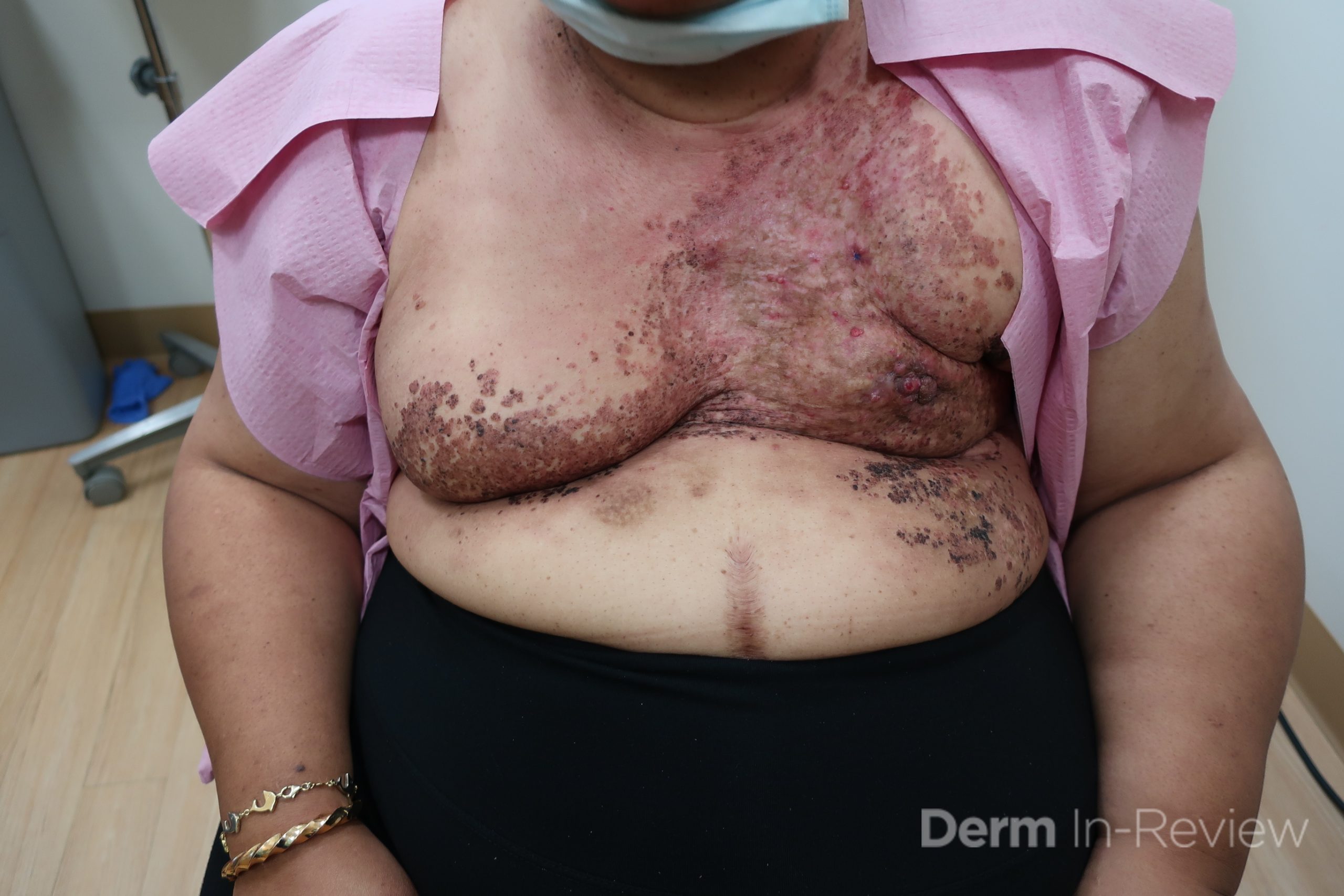
Patient is a 54 year old woman who presents with progressive rash on the anterior torso for 3 months, which she describes as slightly tender bumps. She has history of early stage breast cancer on the left breast treated with lumpectomy and radiation, both completed 9 years ago. She denies any other concerning symptoms.

Biopsy of a representative lesion is most likely to show which of the following?

A.) Dermis infiltrated by atypical neoplasm formed by pleomorphic epithelial cells with hyperchromatic nuclei and distinct nucleoli, disposed in columns and glandular-like structures, amid desmoplastic collagen without associated inflammation, positive immunostaining for HER2, estrogen receptor, and progesterone receptor
B.) Atypical hyperchromatic epithelial cells forming crack-like vascular spaces between collagen bundles, positive immunostaining for CD34, CD31
C.) New vessels wrapping around vascular and adnexal structures, positive immunostaining for HHV-8
D.) Dilation of ectatic superficial vessels, loss of adnexa, papillary dermal pallor, lack of lymphoid band
E.) Acanthosis, pseudoepitheliomatous hyperplasia with suprabasilar crypts of eosinophils

Correct answer: (A) Dermis infiltrated by atypical neoplasm formed by pleomorphic epithelial cells with hyperchromatic nuclei and distinct nucleoli, disposed in columns and glandular-like structures, amid desmoplastic collagen without associated inflammation, positive immunostaining for HER2, estrogen receptor, and progesterone receptor

Explanation:

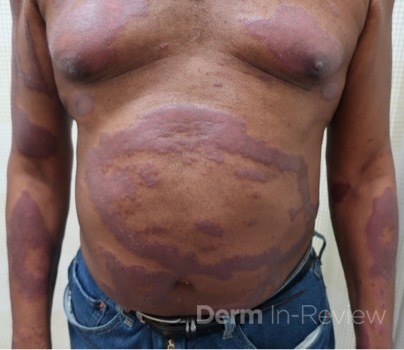
This patient with history of early stage breast cancer who completed surgical and radiation treatment 9 years ago presents with pink to violaceous and hyperpigmented plaques, some studded with pink papules with overlying hemorrhagic crust, predominantly located on the left side of the chest and sprawling out toward the neck and the right chest. Her presentation is most consistent with carcinoma en cuirasse, a presentation of cutaneous metastases most often associated with breast cancer primary. Cutaneous metastases occur in 5-10% of cancer patients and may result from any malignant neoplasm. Prognosis associated with this diagnosis is very poor, as 6 and 12-month mortality rates for these patients are 48% and 64.5%, respectively. Presentations vary, with carcinoma en cuirasse characterized by a diffuse infiltration into the skin, leading to sclerodermoid change. Histopathology may be characterized by a (A) dermis infiltrated by atypical neoplasm formed by pleomorphic epithelial cells with hyperchromatic nuclei and distinct nucleoli, disposed in columns and glandular-like structures, amid desmoplastic collagen without associated inflammation. Positive immunostaining for HER2, estrogen receptor, and progesterone receptor would be suggestive of breast cancer primary. Other presentations of cutaneous metastases include: dermal or subcutaneous nodules, carcinoma erysipeloides, carcinoma telangiectodes, Paget’s disease, alopecia neoplastica, and pyogenic granuloma-like lesions.

Lymphangiosarcoma is high on the differential diagnosis for this patient’s presentation, and may present with non-healing eschars and multiple violaceous nodules. Stewart-Treves syndrome is a rare form of angiosarcoma which arises in the setting of chronic lymphedema, most commonly described in breast cancer patients who have undergone axillary lymph node dissection. Histopathology for this condition may show (B)       atypical hyperchromatic epithelial cells forming crack-like vascular spaces between collagen bundles, with positive immunostaining for CD34, CD31. Patients usually have history of radical mastectomy, as opposed to lumpectomy, and lesions most commonly occur on the ipsilateral upper extremity overlying specific regions of lymphedema, as opposed to widespread lesions occurring over the torso.

Histopathology showing (C) new vessels wrapping around vascular and adnexal structures, with positive immunostaining for HHV-8 would be characteristic of Kaposi sarcoma (KS). There are 4 types of KS, which results directly from human herpesvirus-8 (HHV-8) infection. The classic type usually affects male descendants from the Mediterranean and middle European areas, as well as men in Sub-Saharan Africa, and is not associated with immunodeficiency. Human immunodeficiency virus (HIV)-associated KS is associated with severe immunodeficiency secondary to HIV infection. Endemic KS arises in some parts of Africa, usually in younger patients. Iatrogenic KS results from drug-induced immunosuppression. Presentation classically consists of red to violaceous macules, plaques, or nodules localized anywhere on the skin or mucous membranes, and may even involve the lymph nodes or viscera. There are numerous diverse morphologies reported in the literature, however, including generalized lymphadenopathic, exophytic, disseminated cutaneous and visceral, telangietatic, keloidal, ecchymotic, and localized nodular, plaque, or patch. In this patient who is otherwise well, has no history of immunosuppression, has no known ancestry from any of the aforementioned regions, these widespread tender lesions are more likely representative of cutaneous metastases.

Histopathology showing (D) dilation of ectatic superficial vessels, loss of adnexa, papillary dermal pallor, and lack of lymphoid band would be characteristic of chronic radiation dermatitis. Radiation dermatitis results from direct exposure to radiotherapy delivered by way of external beam ionizing radiation. Acute radiation dermatitis may present as erythema, desquamation, skin necrosis, and ulceration in areas of skin that have been irradiated. Chronic radiation dermatitis may occur up to 10 years or more after the beginning of radiotherapy and involves telangiectasia, fragile skin, and loss of follicular structures. In addition to the current patient’s distinctive morphology suggestive of cutaneous metastases, her radiation was 9 years ago and her eruption is very widespread, making radiation dermatitis much less likely.

Histopathology showing (E) acanthosis, pseudoepitheliomatous hyperplasia with suprabasilar crypts of eosinophils would be suggestive of pemphigus vegetans. Pemphigus vegetans is a localized form of pemphigus vulgaris. Early lesions may present as flaccid pustules, leading to erosions and vegetative and papillomatous plaques usually involving intertriginous areas, scalp, and face. There are 2 predominant subtypes, with the Hallopeau type consisting of more benign disease with longer periods of remission, and the Neumann type involving more disease relapses and requiring higher doses of corticosteroids. Similar to other forms of pemphigus vulgaris, patients have autoantibodies to desmoglein 3 predominantly. This current patient’s lesion morphology and areas of involvement make pemphigus vegetans an unlikely diagnosis.

References

  1. Bolognia JL, Schaffer JV, Duncan KO, Ko CJ. Dermatology essentials E-book. Elsevier Health Sciences; 2014 Feb 26.
  2. Elston D, Ferringer T, Ko CJ, Peckham S, High WA, DiCaudo DJ. Dermatopathology E-Book. Elsevier Health Sciences; 2013 Oct 16.
  3. Schoenlaub, P., et al. “Survival after cutaneous metastasis: a study of 200 cases.” Annales de Dermatologie et de Venereologie. Vol. 128. No. 12. 2001.
  4. Oliveira GM, Zachetti DB, Barros HR, Tiengo A, Romiti N. Breast carcinoma en Cuirasse-case report. Anais brasileiros de dermatologia. 2013 Aug;88(4):608-10.
  5. Mahore SD, Bothale KA, Patrikar AD, Joshi AM. Carcinoma en cuirasse: A rare presentation of breast cancer. Indian Journal of Pathology and Microbiology. 2010 Apr 1;53(2):351.
  6. Sharma A, Schwartz RA. Stewart-Treves syndrome: pathogenesis and management. Journal of the American Academy of Dermatology. 2012 Dec 1;67(6):1342-8.
  7. Etemad SA, Dewan AK. Kaposi sarcoma updates. Dermatologic clinics. 2019 Oct 1;37(4):505-17.
  8. Hegedus F, Mathew LM, Schwartz RA. Radiation dermatitis: an overview. International journal of dermatology. 2017 Sep;56(9):909-14.
  9. Razzaque Ahmed A, Blose DA. Pemphigus vegetans: Neumann type and Hallopeau type. International journal of dermatology. 1984 Mar;23(2):135-41.