2023 Case Studies – Derm In-Review

December 2023 Case Study

By 2023 Case Studies

December 2023 Case Study

Adam Rosenfeld, MD1

  1. Department of Dermatology, George Washington University School of Medicine and Health Sciences

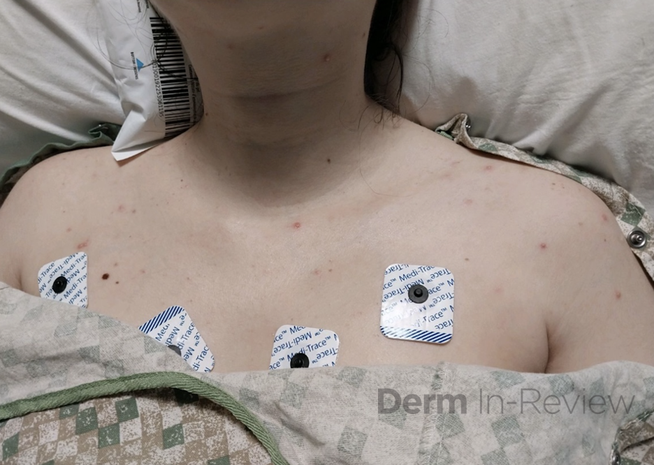
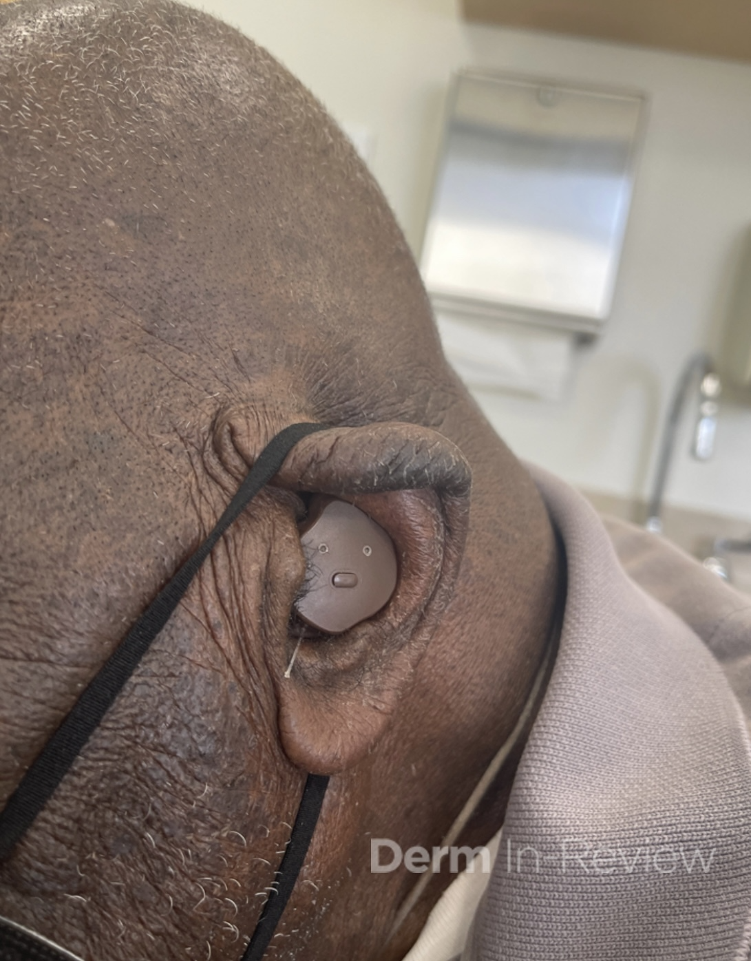
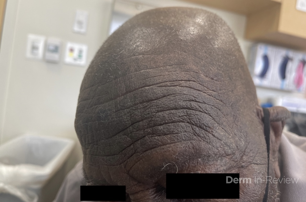
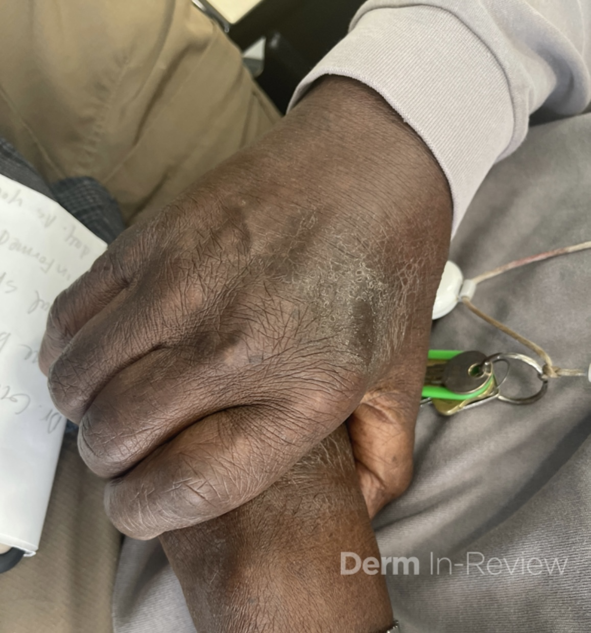
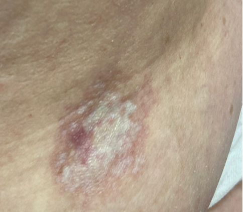
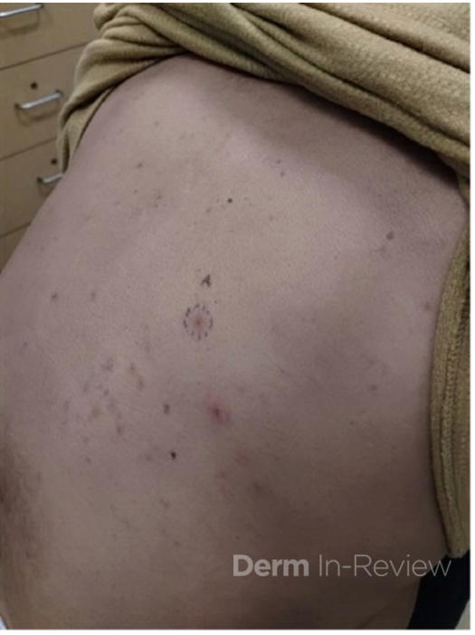
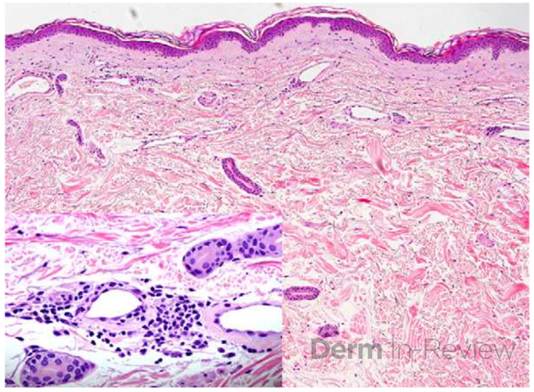
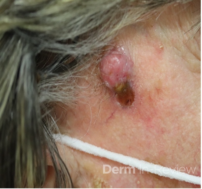
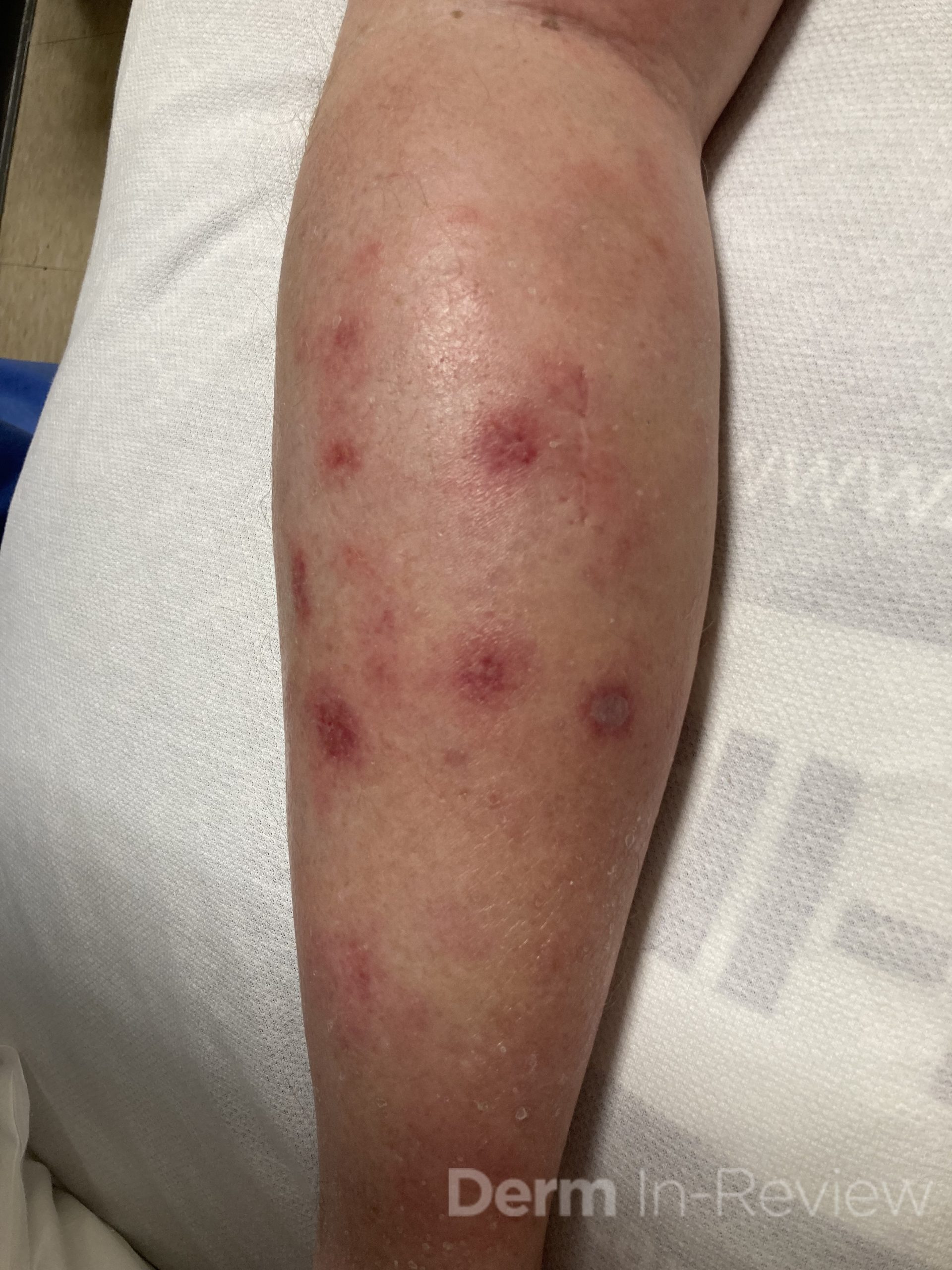
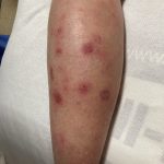
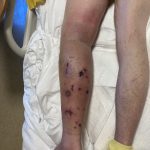
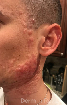
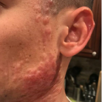
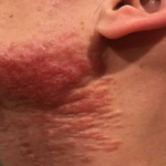
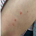
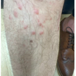
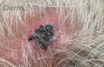
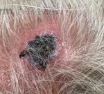
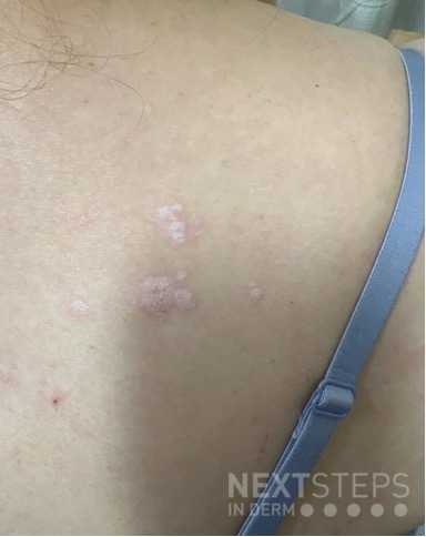
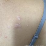
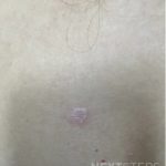
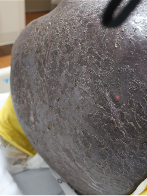
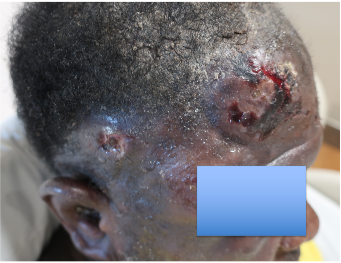
A 46-year-old male with reported history of atopic dermatitis presented to the ED with a 2–3 month history of a diffuse skin eruption as well as multiple growths on his forehead. Review of systems was notable for fatigue and chills. Patient endorsed unintentional weight loss and multiple episodes of night sweats throughout this time frame. On examination, the patient was erythrodermic with diffuse hyperpigmented and erythematous scaly plaques as well as multifocal, somewhat necrotic appearing nodules on the right forehead/temple area (figures 1,2). A punch biopsy demonstrated a superficial to deep dermal infiltrate of atypical lymphoid cells with limited epidermal involvement.

Given the overall presentation, loss of what T-cell marker would be most consistent with the likely diagnosis?

A.) CD20
B.) CD3
C.) CD4
D.) CD7
E.) CD45


.         

Correct Answer:  D

Explanation/Literature review:

Erythroderma and multifocal nodules in the context of unintentional weight loss and constitutional symptoms, are most concerning for Sezary syndrome, which classically demonstrates aberrant loss of CD71. Sezary Syndrome is an aggressive subtype of cutaneous T-cell lymphoma (CTCL) that has been defined by erythroderma, superficial lymphadenopathy, and atypical T-cells in the blood1. Mycosis fungoides (MF), the most common form of CTCL, where disease is typically skin limited, patients often have a much more indolent presentation. Sezary patients on the other hand, present much more acutely and have a poorer prognosis3. The erythroderma in these patients is intensely pruritic1. Patients can also demonstrate diffuse alopecia, keratoderma, eyelid edema and dystrophic nails1. Mycosis fungoides can be difficult to diagnosis and often requires multiple skin biopsies and can mimic atopic dermatitis (AD). Our patient had a known diagnosis of AD, so it is difficult to know if this was CTCL that simply hadn’t been diagnosed yet. The prior school of thought indicated that mycosis fungoides and Sezary syndrome were on a spectrum, where Sezary was progression of a patient’s known disease1. However, while the progenitor cells in MF and Sezary both stem from memory T-cells, in MF they are considered skin resident as opposed to central resident memory T-cells in Sezary2.

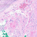
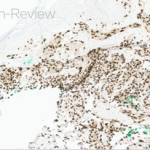
Histologic findings can range from superficial perivascular lymphocytic infiltrate, eosinophilic dermatitis with or without spongiosis, to a lichenoid infiltrate. Pautrier micro-abscesses (collections of atypical lymphocytes within the epidermis) and epidermotrophism (lymphocytes moving from the dermis into the epidermis) can also be seen3,4. Given the variability and often subtilty of histologic findings, the staining pattern is most diagnostic. The infiltrate will be CD3+ and CD4+ (answer choices B and C) but will show aberrant loss of CD7, a marker of immature T-lymphocytes (answer choice D)1. CD20 and BCL2 (answer choice A and E) would be helpful in diagnosing a B-cell lymphoma but would not be relevant in this case4. Although skin involvement is supportive of the diagnosis, to meet criteria for Sezary syndrome, patients most have an atypical, clonal T-cell proliferation within the blood. This can be identified with flow cytometry as well as T-cell rearrangement studies. The population of T-cells must have one of the following: (1) CD4+/CD8+ ratio of >10 (2) CD4+CD7- >40% or CD4+CD26- >301. Further work up includes but is not limited to CBC, CMP, peripheral blood smear and LDH, which can have prognostic importance1. Imaging is indicated to assess for lymphadenopathy as well as visceral involvement (splenomegaly, hepatomegaly)1. Importantly, Sezary patients are inherently immunosuppressed given the abnormal T-cell population as well as barrier dysfunction as it relates to erythroderma2. Sezary syndrome is deemed stage IV from skin involvement but based on lymph node and visceral involvement, more exact staging is done to best guide treatment options, with the help of our oncology colleagues5. Skin directed therapy includes topical or systemic steroids as well as phototherapy or electron beam radiation. Given blood involvement in Sezary syndrome, treatment is typically systemic1. For patients without visceral involvement, therapeutic options include extracorporeal photopheresis, oral retinoids (Bexarotene, Acitretin), interferons or histone deacetylase inhibitors (Vorinostat, Romidespin)1. Some of the targeted therapies include Brentuximab (monoclonal anti-CD30 antibody), mogamulizumab (CCR4 chemokine antibody) as well as Alemtuzumab (monoclonal anti-CD 52 antibody)1. Stem cell transplant is considered the only potentially curative option1.

Incorrect stains in answer choices3

CD20 (choice A) – B-cell antigen. Positive in B-cell lymphomas and negative in T-cell lymphomas. Target for Rituximab.

CD3 (choice B) – Pan T-cell marker. Positive in T-cell lymphomas but negative in B-cell lymphomas

CD4 (choice C) – T-helper lymphocyte marker.

BCL2 (choice E) – An oncogene that inhibits apoptosis. Use in differential diagnosis of B-cell lymphoproliferative disorders.


References:

  1. Sezary syndrome – statpearls – NCBI bookshelf. (n.d.) https://www.ncbi.nlm.nih.gov/books/NBK499874/
  2. Miyashiro D, Souza BCE, Torrealba MP, Manfrere KCG, Sato MN, Sanches JA. The Role of Tumor Microenvironment in the Pathogenesis of Sézary Syndrome. Int J Mol Sci. 2022 Jan 15;23(2):936. doi: 10.3390/ijms23020936. PMID: 35055124; PMCID: PMC8781892.
  3. Elston, D. M. (2019). Dermatopathology. Elsevier.
  4. Sézary syndrome. Pathology Outlines – Sézary syndrome. (n.d.). https://www.pathologyoutlines.com/topic/lymphomanonBsezary.html
  5. Olsen EA, Whittaker S, Kim YH, Duvic M, Prince HM, Lessin SR, Wood GS, Willemze R, Demierre MF, Pimpinelli N, Bernengo MG, Ortiz-Romero PL, Bagot M, Estrach T, Guitart J, Knobler R, Sanches JA, Iwatsuki K, Sugaya M, Dummer R, Pittelkow M, Hoppe R, Parker S, Geskin L, Pinter-Brown L, Girardi M, Burg G, Ranki A, Vermeer M, Horwitz S, Heald P, Rosen S, Cerroni L, Dreno B, Vonderheid EC; International Society for Cutaneous Lymphomas; United States Cutaneous Lymphoma Consortium; Cutaneous Lymphoma Task Force of the European Organisation for Research and Treatment of Cancer. Clinical end points and response criteria in mycosis fungoides and Sézary syndrome: a consensus statement of the International Society for Cutaneous Lymphomas, the United States Cutaneous Lymphoma Consortium, and the Cutaneous Lymphoma Task Force of the European Organisation for Research and Treatment of Cancer. J Clin Oncol. 2011 Jun 20;29(18):2598-607. doi: 10.1200/JCO.2010.32.0630. Epub 2011 May 16. PMID: 21576639; PMCID: PMC3422534.

November 2023 Case Study

By 2023 Case Studies

November 2023 Case Study

Author: Alexis E. Carrington, MD
Reviewed by Karl Saardi, MD

A 30 year old female with a history of ulcerative colitis on ustekinumab and prednisone presents to the hospital with a sudden pustular eruption on the face, chest and arms. She also reports painful ulcers and sores on the tongue, lips and gums. Exam shows scattered pustules and tense vesicles with surrounding erythema on the face, upper trunk and arms (Image 1). There are also punched out ulcerations with a white-yellow base on the mucosal lip and gingiva. Labs are significant for ALT and AST elevated to 1847 units/L and 1163 units/L, respectively.

Which of the following next steps will help with diagnosis?

A.) Obtain an HSV/VZV PCR swab
B.) Obtain an enterovirus PCR swab
C.) Obtain a bacterial culture swab
D.) Obtain a fungal culture swab


This patient has disseminated herpesvirus infection complicated by fulminant hepatitis. This was ascertained by a negative HSV IgG 1 and 2 as well as positive HSV2 PCR from cutaneous swab and blood PCR. There was also a component of an acneiform eruption due to chronic steroid use, resulting in a pustular eruption. Her skin findings and hepatitis improved with initiation of IV acyclovir.

Disseminated HSV is a potentially fatal infection that can have visceral involvement. Organs that can be involved include the liver (HSV hepatitis), the central nervous system (meningoencephalitis), lungs (pneumonitis), bone marrow (neutropenia or thrombocytopenia) or the GI tract (necrotizing enterocolitis).1-3 Disseminated HSV is more common in immunocompromised patients, however can occur in immunocompetent patients. In fact, HSV hepatitis should be considered in the setting of acute fulminant liver failure of unknown etiology, as was the case in this patient.4

An HSV/VZV cutaneous swab for PCR (Answer A) is sensitive and specific for detecting herpesvirus infection, which classically presents as painful tense vesicles and punched out mucosal ulcerations and can cause fulminant hepatitis as a sequelae. Swabs for HSV culture are less sensitive but are needed if testing for acyclovir resistance is required. Coxsackievirus, bacterial and fungal etiologies are less likely to cause this cutaneous and systemic presentation (Answers B-D).

References:

  1. Bolognia, Jean L. Dermatology volume 2. Mosby, 2018.
  2. Srinivasan D, Kaul CM, Buttar AB, Nottingham FI, Greene JB. Disseminated Herpes Simplex Virus-2 (HSV-2) as a Cause of Viral Hepatitis in an Immunocompetent Host. Am J Case Rep. 2021 Aug 3;22:e932474. doi: 10.12659/AJCR.932474. PMID: 34341324; PMCID: PMC8349572.
  3. Fatahzadeh M, Schwartz RA. Human herpes simplex virus infections: epidemiology, pathogenesis, symptomatology, diagnosis, and management. J Am Acad Dermatol. 2007 Nov;57(5):737-63; quiz 764-6. PubMed ID: 17939933
  4. Wei E, Song P, Tan B, Burgin S, Prasad P.  Disseminated herpes simplex virus in adult. In: Goldsmith LA, ed. VisualDx. Rochester, NY: VisualDx; 2023. URL:https://www.visualdx.com/visualdx/diagnosis/?moduleId=101&diagnosisId=52676. Accessed October 15, 2023.

October 2023 Case Study

By 2023 Case Studies

October 2023 Case Study

Author: Alexis E. Carrington, MD
Reviewed by Karl Saardi, MD

A 94 year old male with a medical history of CKD aphasia related a stroke presents to the clinic for a rash on the hands, face and abdomen. History of the rash is significantly limited due to his aphasia. Chart review showed that he was prescribed fluocinonide ointment for suspected atopic dermatitis with no improvement. The patient lives in a skilled nursing facility. Labs showed chronic normocytic anemia with normal folate and B12 levels and chronically elevated transaminases with AST 156 and ALT 193. On exam, there are lichenified hyperpigmented scaly plaques that are slightly photo distributed. Facial features suggestive of leonine facies. (Images 1-3).

Which of the following next steps will help diagnose the rash?

A.) Obtain serum Zinc level
B.) Obtain serum Vitamin C level
C.) Obtain flow cytometry
D.) Obtain serum Niacin levels
E.) Obtaining a mineral prep


Correct Answer: D.) Obtain serum Niacin levels


Explanation of Correct Answer:

Pellagra is a potentially fatal disease due to a lack of the vitamin niacin (B3) or its precursor tryptophan.  Risk factors include poverty, a diet poor not fortified with niacin (such as maize), alcoholism, carcinoid syndrome and drugs such as isoniazid and azathioprine.1,2The rash is typically a symmetrical photo-distributed rash with a thick scale likened to shellac.

Cheilitis and glossitis are commonly associated with pellagra, as well as other vitamin deficiencies. Dementia and diarrhea are other commonly associated symptoms, completing the “4 Ds” of Pellagra (Dementia, Diarrhea, Dermatitis and Death).2 The World Health Organization recommends treatment of pellagra with Nicotinamide 300 mg by mouth divided 2-3 times daily for 3-4 weeks.4 Repletion with a daily multivitamin can be done to treat for other suspected concomitant vitamin deficiencies.

Answer D is correct to confirm a diagnosis of Pellagra. Acrodermatitis enteropathica due to Zinc deficiency commonly involves the peri-oral, acral and intertriginous areas. (Answer A). Vitamin C deficiency can present as perifollicular purpura, gingival bleeding and corkscrew hair (Answer B). Answer C is to evaluate for Sezary syndrome, which can present with leonine facies, however the plaques and tumoral lesions are typically in photo protected areas. Finally a mineral prep would evaluate for scabies, which less likely involves the face and scalp (Answer E). The rest of the answers are reasonable next steps to narrow down the differential diagnosis; however the photo distribution is a typical finding in pellagra.

References

  1. Bolognia, Jean L. Dermatology volume 2. Mosby, 2018.
  2. Hegyi J, Schwartz RA, Hegyi V. Pellagra: dermatitis, dementia, and diarrhea. Int J Dermatol. 2004 Jan;43(1):1-5. doi: 10.1111/j.1365-4632.2004.01959.x. PMID: 14693013.
  3. World Health Organization. Pellagra and its prevention and control in major emergencies. 2000

September 2023 Case Study

By 2023 Case Studies

September 2023 Case Study

by Sapana Desai, MD & Adam Rosenfeld, MD

A 63-year-old woman with history of Hashimoto’s disease, diabetes mellitus, hypertension, and deep venous thrombosis presents with a progressive, multifocal, “rash” involving the extremities including the thighs and buttocks for four-to-five years. Lesions gradually advanced to affecting flexural aspects of the inguinal folds, abdomen, inframammary folds, and axillae. More recently, the patient recognized significant worsening and complains of severe pruritus, pain, and generalized discomfort. Physical examination findings are shown in Figure 1.

Which of the following diagnosis is the patient at an increased risk of developing?

A) Scleromyxedema
B) Interstitial Granulomatous Dermatitis
C) Vulvular Squamous Cell Carcinoma
D) Pheochromocytoma
E) Dermatomyositis


Correct Answer: (C) Vulvular Squamous Cell Carcinoma


Explanation of Correct Answer:

This patient’s clinical presentation, which includes multiple white atrophic plaques comprised of several flat-topped coalescing papules with maceration and scale crust, and a subsequent 4mm punch biopsy revealing a compact stratum corneum, atrophic epidermal with effaced rete, a band-like infiltrate, and associated dermal edema is indicative of extragenital Lichen Sclerosus et Atrophicus (LSetA) with vulvovaginal involvement.

LSetA is an underdiagnosed, chronic, inflammatory mucocutaneous autoimmune dermatosis with increased predilection for the anogenital sites (80% -to- 98%), but can also arise simultaneously in extragenital sites in 15% to 20% of patients- favoring the upper trunk and proximal limbs. Postmenopausal women are predominantly affected and, to a lesser extent, men, prepubertal children, and adolescents, with female-to-male ratios varying between 6:1 and 10:1.1-3 While exact etiology and pathogenesis of LSetA remains elusive, underlying genetics and family predisposition are contributory in 10% of all cases; immunological changes involving increased levels of Th1-specific cytokines, dense T cell infiltration, enhanced BIC/miR expression, as well as autoantibodies against extracellular matrix protein 1 and BP180 antigen also play integral roles. LSetA is characterized by skin atrophy and hypopigmentation, and most commonly incites clinical manifestations of severe pruritus, pain, burning, and sexual dysfunction.2,3 Its relapsing and cumbersome nature warrants every case of LSetA to be treated and managed early, as disease evolvement can lead to scarring and loss of genital anatomical architecture. Furthermore, vulvular lichen sclerosus may evolve to Vulvular Squamous Cell Carcinoma (C) in affected areas with an estimated 5% lifetime risk; however, its association with penile squamous cell carcinoma is not clear.1,3

It is imperative to note that squamous cell carcinoma develops in connection with genital, not extragenital LSetA. Literature suggests that p53 oncogenes, chronic inflammation and oxidative DNA damage are responsible for such malignant transformations. Nonetheless, the risks are significantly decreased by long-term maintenance treatment including an array of topical corticosteroids, topical calcineurin inhibitors, and well as systemic regimens for refractory cases.1,3


Explanation of Incorrect Answers:

Scleromyxedema (A) is a rare chronic cutaneous mucinosis of unknown etiology, often exhibiting association with rheumatologic diseases and monoclonal gammopathy (IgG-lambda). The disease affects adults between 30 -to- 70 years of age, with no gender predilection. Patients experience widespread symmetric eruptions of waxy, firm erythematous papules measuring 2 -to- 3cm diameter which can present close grouping in a linear distribution.1,4 Gradually, papules evolve to hardened plaques, showing marked sclerosis. In addition to cutaneous changes, 90% of patients experience plasma cell dyscrasia, in conjunction with multisystemic manifestations that may involve cardiovascular, gastrointestinal, respiratory, skeletal, muscular, renal, and nervous systems, resulting in significant morbidity and mortality. Histologically, scleromyxedema is characterized by excessive dermal mucin deposition, fibroblast proliferation, and fibrosis; still, histological findings in more than 20% of afflicted patients differs from the classic triad showing lesions that resemble interstitial granuloma annulare.4 Scleromyxedema is an unpredictable, progressive, and debilitating disease, and its relative rarity and elusive pathophysiology has precluded consensus on any one optimal treatment regimens. Even so, current literature supports aggressive treatment with high-dose intravenous immunoglobulins (IVIG) is promising while inciting the least number of side effects.

Interstitial Granulomatous Dermatitis (B) is an infrequent dermatosis that most commonly affects females, and is typically seen in the setting of rheumatic diseases [e.g. rheumatoid arthritis], but also hematological disorders, internal malignancies, infections, and antihypertensive drugs.5 Clinically, the disease is characterized by multiple erythematous papules and plaques, often with annular configuration, predominantly involving inner aspects of the limbs, lateral trunk, and intertriginous sites.5,6 The presence of cord-like indurated lesions (“rope sign”) is often considered pathognomonic. Uniquely, the histopathological examination defines the diagnosis, with presentation of dense and diffuse interstitial infiltrates within the reticular dermis- composed of histocytes in a palisade arrangement and sometimes with necrobiosis of collagen, and deposition of mucin with absence of both vasculitis and neutrophilia.1,5,6

Pheochromocytoma (D) may be associated with neurofibromatosis type 1- an autosomal dominant inherited neurocutaneous disorder due to mutations in NF1 gene on 17q11.2. Pheochromocytoma is a rare neuroendocrine catecholamine-producing tumor of the adrenal medulla that synthesizes and stores excessive amounts of norepinephrine and epinephrine, which when released can evoke life-threatening cardiovascular complications.8 It may be classified as sporadic or familial, with the latter accounting for 10% of all pheochromocytoma cases, and exhibits strong association with hereditary inheritance of multiple endocrine neoplasia IIA and IIB, neurofibromatosis type 1, tuberous sclerosis, von Hippel-Lindau syndrome, Sturge-Weber syndrome, and simple familial pheochromocytoma.8,9 Up to 40% of pheochromocytomas are associated with known genetic mutations, with the most common encompassing NF-1 gene, RET proto-oncogene, VHL gene, and genes encoding succinate dehydrogenase subunits.8,9

Dermatomyositis (E) is an inflammatory myopathy with autoimmune pathogenesis affecting women more commonly than men, with a bimodal peak of incidence between ages 5 and 14 and ages 45 and 65. Clinical presentation classically encompasses a heliotrope rash (periorbital erythema with edema, occasionally involving the cheeks and nose), Holster sign (symmetric poikiloderma of the lateral thighs below the greater trochanter), and V-sign (confluent erythematous patches over the upper central chest and lower anterior neck).1,7  Dermatomyositis is sub-classified into adult-onset disease and juvenile disease (JDM). Serum antinuclear autoantibodies are often present, as are other myositis-specific autoantibodies, which are useful as prognostic indicators while aiding in diagnosis and management of disease. Dermatomyositis is related to and possibly results from an immune-mediated process triggered by outside factors like drugs, infectious agents, and malignancy in individuals with genetic predisposition.7

References

  1. Bolognia, J., Schaffer, J., & Cerroni, L. (2018). Dermatology (Fourth edition.). Philadelphia,Pa: Elsevier. of clinical medicine. Retrieved August 11, 2022, from https://pubmed.ncbi.nlm.nih.gov/32516921/
  2. Elston, D. M. (2019). Dermatopathology. Elsevier.
  3. Krapf JM, Mitchell L, Holton MA, Goldstein AT. Vulvar lichen Sclerosus: current perspectives. Int J Women’s Health. (2020) 12:11–20. doi: 10.2147/IJWH.S191200
  4. Koronowska, S., Osmola-Mankowska, A., Jakubowicz, O., Zaba, R. Scleromyxedema: a rare disorder and its treatment difficulties. Advances in Dermatology and Allergology/Postepy Dermatol Alergol. (April 2013); 30(2): 122-126.
  5. Veronez, I., Luiz Dantas, F., Valente, N., Kakizaki, P., Yasuda, T., Cunha, Thais. Interstitial granulomatous dermatitis: rare cutaneous manifestation of rheumatoid arthritis. Anais Brasileiros De Dermatologia. (May 2015); 90(3): 391-393
  6. Coutinho, I., Pereira, N., Gouveia, M., Cardoso, J., Tellechea, O. Interstitial Granulomatous Dermatitis: A Clinicopathological Study. The American Journal of Dermatopathology. (August 2015)); 37(8): 614-619.
  7. DeWane ME, Waldman R, Lu J. Dermatomyositis: clinical features and pathogenesis. Journal of the American Academy of Dermatology. 2020 Feb 1;82(2):267-81.
  8. Zografos, G., Vasiliadis, G., Zagouri, F., Aggeli, C., Korkolis, D., Vogiaki, S., Pagoni, M., Kaltsas, G., Piaditis, G. Pheochromocytoma Associated with Neurofibromatosis Type 1: Concepts and Current Trends. World Journal of Surgical Oncology. 2010; 8(14).
  9. Naranjo, J., Dodd, S. MD, Martin, Y. MD. Perioperative Management of Pheochromocytoma. Journal of Cardiothoracic and Vascular Anesthesia. 2017; 31(4): 1427-1439.

August 2023 Case Study

By 2023 Case Studies

August 2023 Case Study

by Nidhi Shah, MD

A 67-year-old male with past medical history of hypertension, type 2 diabetes, cirrhosis, and psoriasis presented for a pruritic rash involving his scalp, trunk, and extremities for a year. On examination, patient had small erythematous macules and telangiectasias scattered on the trunk, extremities, and scalp (Figure 1). A punch biopsy of the left lower back was obtained (Figure 2)1. Laboratory findings demonstrated an elevated tryptase level.

Based on the clinical presentation and biopsy and laboratory findings, what is the next best test?

A.) CK
B.) Hepatitis panel
C.) c-KIT
D.) TSH
E.) HIV


Correct Answer: C.) c-KIT


Explanation of Correct Answer:

Patient’s telangiectatic and macular rash with biopsy findings of mononuclear infiltrate of mast cells around the capillaries in the upper dermis raises concerns for telangiectasia macularis eruptive perstans (TMEP), a rare form of cutaneous mastocytosis. TMEP most commonly manifests in adulthood, although rare reports of pediatric cases have been reported.1,2 Although previously considered to be a solely cutaneous disease, recent evidence suggests the presence of systemic involvement including bone marrow, gastrointestinal tract, liver, spleen and lymph node.2,3

TMEP classically presents as small, irregular red-brown blanchable telangiectatic macules ranging from 2-4mm in diameter overlying a tan background.2 These lesions generally are non-pruritic and involve the trunk and proximal extremities in a symmetric pattern, sparing the palms, soles, and face in most cases. Commonly positive in urticaria pigmentosum or maculopapular cutaneous mastocytosis (UP/MPCP), Darier’s sign is usually negative or slight in TMEP given the low number of mast cells involved.2

If characteristic skin lesions are accompanied by symptoms like flushing, diarrhea, dyspnea, tachycardia, pruritis, syncope, or indications of anaphylaxis, it’s important to consider the possibility of systemic involvement.2 Serum tryptase and 24-hour urine histamine levels are used as preliminary test to investigate for systemic involvement with tryptase levels >20 µg/L concerning for systemic mastocytosis.2 In adults, a c-KIT proto-oncogene mutation is most commonly observed which leads to mast cell hyperplasia.4 The type III transmembrane tyrosine kinase receptor c-kit has an extracellular domain that binds to mast cell growth factor and activation leads to mast cell growth and production.2 Although the mutation is not present in all patients, proto-oncogene alternations may be correlated with more aggressive systemic involvement, making it important to check for the mutation.4

There is no gold standard treatment for TMEP and treatment is based upon the type of systemic involvement and clinical symptoms. Lifestyle modifications to avoid stimulation of mast cell degranulation such as exposure to sunlight and extreme temperatures, alcohol, and drugs should be practiced. Additional treatment modalities include antihistamines, PUVA, and narrow-band UVB.2,4 Beyond management of symptoms, staging studies are required to evaluate for aggressive disease. The only FDA approved treatment for cytoreduction in mastocytosis is avapritinib, an oral small molecule inhibitor of D816V-mutant KIT.5


References

  1. Martín-Fuentes A, Pastor-Nieto MA, De Eusebio-Murillo E. Telangiectatic Macules in a 90-Year-Old Woman. Actas Dermo-Sifiliográficas (English Edition). 2012;103(1):65-66. doi:10.1016/j.adengl.2011.07.006
  2. Watkins CE, Bokor WB, Leicht S, Youngberg G, Krishnaswamy G. Telangiectasia Macularis Eruptiva Perstans: more than skin deep. Dermatol Reports. 2011;3(1):e12. doi:10.4081/dr.2011.e12
  3. Severino M, Chandesris MO, Barete S, et al. Telangiectasia macularis eruptiva perstans (TMEP): A form of cutaneous mastocytosis with potential systemic involvement. J Am Acad Dermatol. 2016;74(5):885-891.e1. doi:10.1016/j.jaad.2015.10.050

June 2023 Case Study

By 2023 Case Studies

June 2023 Case Study

by Sapana Desai, MD & Kamaria Nelson, MD

A 93-year-old Caucasian female patient with significant past medical history of atrial fibrillation, chronic obstructive pulmonary disease, and rheumatoid arthritis presents with a single, painless, cherry red colored, glistening dome-shaped nodule on her right mid temporal scalp that appeared two months ago. Although asymptomatic, the palpable nodule rapidly evolved to measuring 3.5 x 4.5 x 4cm. Physical examination findings are shown in Figure 1.

Which of the following immunohistochemical marker is strongly associated with the patient’s diagnosis?

A) HHV8 LNA-1
B) CK20
C) Factor XIIIa
D) PHLDA1
E) CD31


Correct Answer: B.) CK20


Explanation of Correct Answer:

This elderly patient’s clinical presentation, which entails spontaneous emergence of a rapidly growing, asymptomatic reddish to violaceous spherical tumor with a smooth, shiny surface and a soft to turgid elastic consistency localized to the face- a region with greatly increased ultraviolet (UV) exposure, is highly suspicious for Merkel Cell Carcinoma (MCC).

MCC is a rare and aggressive cutaneous malignancy of neuroendocrine origin that most frequently presents with head and neck distribution and is characterized by high rates of local recurrence and nodal metastasis; five-year survival rates of all stages are 50-to-60%.1 MCC arises in individuals of advanced age with peak incidence of 75 years of age, and predominantly affects Caucasian males when compared to women and other ethnic groups. While etiology of MCC remains elusive, clonal integration of the Merkel cell DNA double-stranded polyomavirus (MCPyV) into the host genome, or UV-mediated genetic aberrations affecting the tumor suppressor p53 and RB1 pathways as well as epigenetic factors that activate oncogenes, are accepted theories in carcinogenesis.1,2,3 Other culprit risk factors encompass chronic UV exposure, lymphoproliferative diseases, and iatrogenic or pathological immunosuppression.3

Histopathologic assessment of MCC will reveal infiltration of the dermis and subcutaneous tissue- often sharing a connection with overlying epidermis and adnexal tissue, and exhibit sheets and nests of crowded basaloid cells with a finely granular “salt and pepper” chromatic pattern, indistinct nucleoli, and scant cytoplasm.1 Mitoses and extensive necroses are common and reflect the detrimental nature of these tumor. Because histomorphology of MCC is rarely specific, immunohistochemistry is imperative for definitive diagnosis and can be confirmed via detection of CK20 (B), typically identified in perinuclear granules; metastasis of Small Cell Lung Carcinoma (SCLC) can be excluded if thyroid transcription factor 1 (TTF-1) is negative. In unusual circumstances if staining patterns for MCC are atypical or CK20 fails to be expressed, additional immunohistochemical markers including Pancytokeratin (AE1/3), chromogranin A, neuron specific enolase (NSE), synaptophysin, INSM1, or N-CAM (CD56) may be used.1,4

Surgical modalities are most often utilized for primary MCC neoplasms, whether it be via wide local excisions (WLE) with 1-to-2cm margins, Mohs micrographic surgery (MMS), or excision with complete circumferential peripheral and deep margin assessment. In non-excisable circumstances and one-third of MCC cases with distant nodal and intra-abdominal metastases, immunotherapy with checkpoint inhibitors targeting programmed death receptor-1 (PD-1)/ programmed dead ligand-1(PD-L1) pathways have proved to be beneficial, preventing inhibition of T-cells and leading to increased immune system activity and antitumor responses. Current marketed immunotherapies for advanced stages of MCC include: Avelumab– a humanized anti-PD-L1 antibody (dose: 800mg IV q2weeks), Pembrolizumab– a humanized IgG4 antibody directed against PD-1 (dose: 2mg/kg q3weeks x 3 years), and Nivolumab- a fully human IgG4 antibody directed against PD-1 (dose: 240mg IV q2weeks).15


Explanation of Incorrect Answers:

Human Herpes Virus-8 Latency Associated Nuclear Antigen 1 (HHV8 LNA-1) (A) immunohistochemistry positivity is both sensitive and highly specific for diagnosis of Kaposi Sarcoma (KS), an angioproliferative mesenchymal malignant neoplasm caused by infection with Kaposi Sarcoma-Related Herpesvirus.5,6 Expression of HHV8 LNA-1 causes binding of p53 and suppression of apoptosis. Subsequent activation of NF-kB elicits up-regulation of VEGF and bFGF and consequent neoangiogenesis, prompting characteristic KS histomorphological features of dermal masses of spindle cells with extravasated red blood cells and cellular atypia.5 KS is classified into four epidemiological subtypes: 1) classic form- presenting increased predilection amidst elderly men of Mediterranean and Eastern European descent with lower extremity involvement, 2) endemic African form– occurring amongst pediatric populations with generalized lymph node involvement, 3) AIDS-related form- afflicting HIV positive male homosexuals with diffused dermal and systemic involvement, and 4) iatrogenic form- affecting immunosuppressed individuals with analogous dissemination and effects as the latter.5,6 While cutaneous findings in KS range from scattered pink to violaceous patches and papules to rapidly progressive, multicentric, ulcerated plaques and nodules with dissemination to visceral organs [eg. lungs and gastrointestinal system], lesions are often painful with associated lymphedema and secondary infection.6

Factor XIIIa (C) immunohistochemistry is a strongly positive staining marker for Dermatofibromas.7 Regarded as benign fibrohistocytic neoplasms, Dermatofibromas clinically present as asymptomatic, solitary, firm to hard, often hyperpigmented, slow-growing, plaque or cutaneous nodules with or without overlying dermal changes.7,8  The “dimple sign” is a characteristic finding where lateral inward digital pressure of the skin produces central dimpling over the lesion. Dermatofibromas occur in people of all ages with increased predilection for the extremities, and most commonly ensue spontaneously with no inciting event but in a fifth of all reported cases are attributed to a primary reactive process from history of local trauma, such as vaccination or an insect bite.8 Classical histopathological features consist of ill-defined intersecting bundles of spindle cells with collagen trapping.7,8 Several dermoscopic patterns of Dermatofibromas are described, of which the most common is central white scar-like patch with delicate pigmentary network at the periphery; other typical patterns include total homogenous pigmentation and irregular crypts associated with pseudofollicular openings.7,8 It is especially vital to differentiate Dermatofibromas from its similar-appearing, more advanced and aggressive counterpart, Dermatofibrosarcoma protuberans [DFSP], with the latter staining positive for CD-34 and negative for FactorXIIIa.8

PHLDA1 (D) along with stromal CD34 and CD10 immunohistochemistry positivity is often used to delineate Trichoblastomas from Basal Cell Carcinoma as the two diagnoses commonly share histopathological features, including the presence of fissures between the epithelium and spindled-fibroblast follicular stroma, epithelial nests, and cells in a palisading arrangement on the tumor periphery.9,11 Trichoblastomas are rare, benign dermal tumors arising from aberrant proliferation of primitive follicular germinate cells. They clinically present as asymptomatic, solitary, skin-colored, slow-growing nodules that measure less than 2 cm in diameter on the face or scalp. Overlying epidermis is alopecic, thin, and hyperpigmented, and occasionally exhibit superficial telangiectasias and ulcerations.10 Trichoblastomas may emerge sporadically, in association with hereditary disease including Brooke-Spiegler disease and Brooke-Fordyce syndrome, or within a nevus sebaceous, and typically affect adults of both genders within their fifth and sixth decades of life. More often than not, Trichoblastomas have favorable prognosis, with low incidence of progression, recurrence, or association with malignancy.9,10,11

Immunohistochemistry positivity for CD31 (E) as well as for other vascular markers including CD34 and factor VIII antigen are suggestive for diagnosis of pyogenic granuloma, a benign vascular tumor that arises within cutaneous and mucosal tissues affecting patients of all ages.12 Although staining is often unnecessary given the characteristic clinical history and histological architecture, atypical lesions may necessitate its use. Histopathology shows highly vascularized proliferation of granulation tissue, lobular arrangements of capillary vessels and proliferating endothelial cells delineated by fibrous septae, scattered fibroblasts, and a variegated inflammatory infiltrate.12,13 Pyogenic granuloma appears as a solitary, red, pedunculated papule that is greatly friable, and exhibits rapid exophytic growth. Lesions often undergo ulceration and can bleed profusely with even minor trauma.14 While the pathophysiology is not fully elucidated, imbalance of pro-angiogenic and anti-angiogenic factors, hormonal influences with increased estrogen and progesterone levels during pregnancy, reactive granulation tissue from negligible injuries, and iatrogenic use of specific medications are all plausible contributors.12,13,14


References

  1. Becker, JC., Beer, A., DeTemple, V., Eigentler, T., Flaig, M., Gambichler, T., Grabbe, S., Holler, U., Klumpp, B., Lang, S., Pfohler, C., Posch, C., Prasad, V., Schlattmann, P., Ter-Nedden, J., Terheyden, P., Thoms, K., Vordermark, D., Ugurel, S. S2K Guideline- Merkel Cell Carcinoma [MCC, Neuroendrocrine Carcinoma of the Skin]- Update 2022. Journal of the German Society of Dermatology. 2023; 21(3): 305-320.
  2. Hernandez, L., Mohsin, N., Yaghi, M., French, F., Dreyfuss, I., Nouri, K. Merkel Cell Carcinoma: An Updated Review of Pathogenesis, Diagnosis, and Treatment Options. Dermatologic Therapy. March 2022; 35(3).
  3. MD, Y., THakuria, M. MD. Merkel Cell Carcinoma Review. Hematology/Oncology Clinics of North America. February 2019; 33(1): 39-52.
  4. Brady, M., Spiker, A. Merkel Cell Carcinoma of the Skin. 2022.
  5. Biship, B., Lynch, D. Kaposi Sarcoma. 2022
  6. Etemad, S., Dewan, A. Kaposi Sarcoma Updates. Dermatologic Clinics. October 2019; 37(4): 505-517.
  7. Singh, S., Patra, S., Bhari, N. Dermatofibroma Over the Face. Indian Dermatology Online Journal. Jan-Feb 2019; 10(1): 94-95.
  8. Myers, D., Fillman, E. Dermatofibroma. 2022.
  9. Schukow, C., Ahmed, A. Trichoblastoma and Trichoepithelioma. StatPearls. 2022.
  10. Patel, P., Nawrocki, S., Hinther, K., Khachemoune, A. Trichoblastomas Mimicking Basal Cell Carcinoma: The Importance of Identification and Differentiation. Cureus. May 2020; 12(5).
  11. Cazzato, G., Cimmino, A., Colagrande, A., Arezzo, F., Lospalluti, L., Sablone, S., Lettini, T., Resta, L., Ingravallo, G. The Multiple Faces of Nodular Trichoblastoma: Review of the Literature with Case Presentation. Dermatopathology. 2021; 8(3): 265-270.
  12. Wollina, U., Langner, D., Franca, K., Gianfaldoni, S., Lotti, T., Tchernev, G. Pyogenic Granuloma- A Common Benign Vascular Tumor with Variable Clinical Presentation: New Findings and Treatment Options. Open Access Macedonian Journal of Medical Sciences. July 2017; 5(4): 423-426.
  13. Komakech, D., Ssenkumba, B. Pyogenic Granuloma. New England Journal of Medicine. November 2022; 387(21): 1979.
  14. Sarwal, P., Lapumnuaypol, K. Pyogenic Granuloma. 2022.
  15. Gauci, M., Aristei, C., Becker, J. Garbe, J., Lebbe, C. Diagnosis and Treatment of Merkel Cell Carcinoma: European Consensus-Based Interdisciplinary Guideline- Update 2022. European Journal of Cancer. August 2022; 17(1): 203-231.

May 2023 Case Study

By 2023 Case Studies

May 2023 Case Study

by Emily Murphy, MD1, Karl Saardi, MD1

  1. Department of Dermatology, George Washington University School of Medicine and Health Sciences

A 65-year-old male with a past medical history of HIV (CD4 count 250, undetectable viral load) and chronic congestion presented with a chief complaint of recurrent cellulitis of the left lower leg. The redness and swelling persisted for 3 months despite antibiotics. On presentation, he had circumferential edema and induration of the left lower leg with erythematous, tender nodules and early bullae (figure 1). A week later, his disease progressed with ulceration and necrosis of these tender nodules (figure 2). A punch biopsy was done, which showed an atypical lymphoid infiltrate in the dermis and subcutaneous tissue with an angiocentric pattern, composed of large, pleomorphic cells. Extensive necrosis was present with karyorrhetic debris. Immunostains were positive for T cells (CD2), natural killer cells (CD56), and cytotoxicity (granzyme B). A biopsy of the nasal septum showed similar findings. The patient was diagnosed with extranodal NK/T cell lymphoma, nasal type.

Which virus is part of the diagnostic criteria for this lymphoma?

A.) Human herpesvirus-8
B.) Polyomavirus
C.) Human immunodeficiency virus
D.) Ebstein Bar Virus
E.) Cytomegalovirus

Correct Answer: D) Ebstein Bar Virus

Extranodal NK/T cell lymphoma, nasal type is an aggressive, non-Hodgkin lymphoma that has four criteria according to the World Health Organization: vascular damage, prominent necrosis, a cytotoxic phenotype, and association with the Ebstein bar virus (EBV, answer D).1,2 EBV positivity is seen in the both the skin and serum. Pathologically, the atypical lymphocytes are positive for cytotoxic markers (granzyme B, peforin), natural killer cell markers (CD2), T cell markers (cytoplasmic CD3), and EBV.1 Extranodal NK/T cell lymphoma is rare in the United States, but its incidence is increasing. It is more common in East Asia and Latin America.1

This lymphoma typically affects the upper aerodigestive tract, causing congestion, epistaxis, or necrotizing lesions of the nose or hard palate.1 Other sites can be involved, including the extranasal skin, gastrointestinal tract, testes, or lungs.1,3,4 On the skin, either a generalized morbilliform eruption or purpuric nodules with or without ulceration can be seen.3 Hemophagocytic lymphohistiocytosis can occur, but is rare, occurring in only 3% of patients.1

NK/T cell lymphoma has an increased incidence in patients with HIV; a study of 93 patients with this lymphoma found that it occurred at a 15 times higher incidence in patients with HIV compared to the general population.5 However, HIV (choice C) is not part of the diagnostic criteria like EBV. Human herpesvirus-8 (Kaposi sarcoma), Polyomavirus (Merkel Cell Carcinoma), and cytomegalovirus are not associated with Extranodal NK/T cell lymphoma (choices A, B, E).

Treatment is determined by the Ann Arbor Staging and various prognostic tools, like the PINK-E system (age > 60 years, Stage III/IV, nonnasal primary localization, distant lymph node involvement, and detectable plasma EBV DNA).6 Depending on the stage and prognosis, treatment is typically with an asparaginase-based regimen called SMILE (dexamethasone, methotrexate, ifosfamide, L-asparaginase, and etoposide).1,6,7 NK/T cell lymphoma cells are unable to synthesize the amino acid, asparagine, and as this amino acid is depleted with asparaginase, protein synthesis is disrupted and the cells ultimately undergo apoptosis.6 Chemotherapy is typically followed by involved field radiation therapy.1  Various immunotherapies are currently being studied for patients with recalcitrant or relapsed disease as well, including programmed death 1 inhibitors, JAK 3 inhibitors, or anti-CD30 therapies.8 Response to therapy can be monitored with EBV DNA titers, which should decrease with treatment.1

References

  1. Haverkos BM, Pan Z, Gru AA, et al. Extranodal NK/T Cell Lymphoma, Nasal Type (ENKTL-NT): An Update on Epidemiology, Clinical Presentation, and Natural History in North American and European Cases. Curr Hematol Malig Rep. 2016;11(6):514-527. doi:10.1007/s11899-016-0355-9
  2. Willemze R, Cerroni L, Kempf W, et al. The 2018 update of the WHO-EORTC classification for primary cutaneous lymphomas. Blood. 2019;133(16):1703-1714. doi:10.1182/blood-2018-11-881268
  3. Orlowski GM, Tan AJ, Evan-Browning E, Scharf MJ. Primary cutaneous nasal-type NK/T-cell lymphoma presenting as purpuric nodules on the lower leg. JAAD Case Rep. 2020;6(10):1075-1078. doi:10.1016/j.jdcr.2020.08.007
  4. Rahal A, Reddy PS, Alvares C. Extranodal NK/T-Cell Lymphoma, Nasal Type, Presenting as a Breast Mass. Cureus. 7(12):e408. doi:10.7759/cureus.408
  5. Castillo J, Pantanowitz L. HIV-Associated NK/T-Cell Lymphomas: A Review of 93 Cases. Blood. 2007;110(11):3457-3457. doi:10.1182/blood.V110.11.3457.3457
  6. van Doesum JA, Niezink AGH, Huls GA, Beijert M, Diepstra A, van Meerten T. Extranodal Natural Killer/T-cell Lymphoma, Nasal Type: Diagnosis and Treatment. Hemasphere. 2021;5(2):e523. doi:10.1097/HS9.0000000000000523
  7. Yamaguchi M, Kwong YL, Kim WS, et al. Phase II study of SMILE chemotherapy for newly diagnosed stage IV, relapsed, or refractory extranodal natural killer (NK)/T-cell lymphoma, nasal type: the NK-Cell Tumor Study Group study. J Clin Oncol. 2011;29(33):4410-4416. doi:10.1200/JCO.2011.35.6287
  8. Lv K, Li X, Yu H, Chen X, Zhang M, Wu X. Selection of new immunotherapy targets for NK/T cell lymphoma. Am J Transl Res. 2020;12(11):7034-7047.

April 2023 Case Study

By 2023 Case Studies

April 2023 Case Study

by Sapana Desai, MD & Azam Qureshi, MD

A 37-year-old male presents with a two-year history of multiple “bumps” on the face. Asymptomatic, small, pea-sized, skin-colored papules were first noted over his left lateral face and mandibular jawline. Gradually, the papules evolved into plaques, with some appearing as large as 3 centimeters. During this time, several new erythematous papules arose along the left parotid region, right elbow, and right shin, and were noted to be painful to palpation and when exposed to cold temperature. Physical examination findings are shown in Figure 1a-d.

Which of the following is commonly associated with the patient’s diagnosis?

A) Iron Deficiency Anemia
B) Renal Cell Carcinoma
C) Angiomyolipoma
D) Pheochromocytoma
E) Eccrine Spiradenoma


Correct Answer: B) Renal Cell Carcinoma


Explanation of Correct Answer:

This patient’s clinical presentation, which includes multiple erythematous, tender, dermal nodules of varying sizes in both a clustered and scattered distribution along the face and extensor surfaces of the extremities is highly suspicious for piloleiomyomas.1,2

A piloleiomyoma, regarded as the most common variant of cutaneous leiomyomas [CL] followed by angioleiomyoma, and dartoic [genital] leiomyoma, is a benign dermal neoplasm arising from smooth muscles of the erector pilorum muscle and can be solitary or multiple.1,3 Piloleiomyomas emerge with peak incidence between 20 and 40 years of age, and equally affect both genders. 90 percent of afflicted patients experience spontaneous and/or paroxysmal sharp pain often precipitated by light touch, pressure, or cold temperatures, that is believed to be attributed to cutaneous nerve compression, injury, or by contraction of tumor muscle fibers.1,2,3 While pathophysiology of piloleiomyoma remains elusive, underlying autosomal dominant [AD] inheritance of fumerate hydratase [FH] mutations are potentially associated with the development of multiple cutaneous piloleiomyoma in Hereditary Leiomyomatosis and Renal Cell Carcinoma [HLRCC] (B), also known as Reed Syndrome.4

HLRCC is a rare AD familial cancer syndrome caused by a germline amorphic allele of the FH gene, in which susceptible individuals are at increased risk for the development of cutaneous leiomyomas, early onset of multiple uterine leiomyomas, and type 2 papillary renal cell cancer.4,5 Recent meta-analyses suggest approximately 20% to 35% of individuals with germline FH mutations develop RCC and die within 5 years of diagnosis.6 Given the aggressive and metastatic nature of these tumors, annual surveillance in patients with known HLRCC should begin at 10 years of age, and surgical removal of even small sized renal tumors is recommended.4,5,


Explanation of Incorrect Answers:

Iron Deficiency Anemia (A) may be associated with Blue Rubber Bleb Nevus Syndrome [BRBNS/ Bean’s Syndrome]. Patients have characteristic cutaneous findings of multiple blue to violaceous soft and compressible nodules on the skin or mucous membranes that present at birth or in early childhood and develop numerous venous malformations involving the skin and gastrointestinal tract over their’ lifetime.7,8 Typical lesions range in size from a few millimeters to up to 4 centimeters in diameter, are rubbery in consistency, and gradually evolve with time eliciting pain most prevalent at night.8 Uniquely, lesions swell in gravity-dependent positions, and patients exhibit focal areas of hyperhidrosis overlying these lesions. Although most cases of BRBNS are sporadic, autosomal dominant inheritance has been suggested, as well as studies reporting stem cell factor/c-kit signaling systems contributing to vascular overgrowth. Lower gastrointestinal bleeding is the most common symptom ranging from obscure to massive bleeding leading to severe iron deficiency anemia. Other rare complications may include perforation, intussusception, intestinal torsion, disseminated intravascular coagulation, and thrombocytopenia.7,8

Renal Angiomyolipoma (C) is strongly associated with tuberous sclerosis complex [TSC], an AD, neurocutaneous disorder characterized by an increased predisposition to hamartoma formation, caused by genetic mutations for the proteins hamartin and tuberin, TSC1 and TSC2, respectively.9  Clinical presentation of TSC is highly variable given its multisystemic involvement, with epilepsy leading the list of neurological manifestations and accounting for 94% of affected patients, followed by cortical tubors, subependymal nodules, and subependymal giant cell astrocytomas. Frequent cutaneous manifestations encompass ash-leaf shaped hypomelanotic macules, facial angiofibroma’s, and shagreen patches.9,10 Renal angiomyolipomas may emerge in 55% to 75% of TSC patients with various longitudinal studies reporting a 75% prevalence of renal angiomyolipomas by 10.5 years of age.10,11 Although benign, TCS-associated renal angiomyolipomas are bilateral and multifocal, and when larger than 4 centimeters pose greater risks for potentially catastrophic hemorrhage when compared with isolated renal angiomyolipoma.11

Pheochromocytoma (D) may be associated with neurofibromatosis type 1- an autosomal dominantly inherited neurocutaneous disorder due to mutations in NF1 gene on 17q11.2. Pheochromocytoma is a rare neuroendocrine catecholamine-producing tumor of the adrenal medulla that synthesizes and stores excessive amounts of norepinephrine and epinephrine, which when released can evoke life-threatening cardiovascular complications.12 It may be classified as sporadic or familial, with the latter accounting for 10% of all pheochromocytoma cases, and exhibits strong association with hereditary inheritance of multiple endocrine neoplasia IIA and IIB, neurofibromatosis type 1, tuberous sclerosis, von Hippel-Lindau syndrome, Sturge-Weber syndrome, and simple familial pheochromocytoma.12,13 Up to 40% of pheochromocytomas are associated with known genetic mutations, with the most common encompassing NF-1 gene, RET proto-oncogene, VHL gene, and genes encoding succinate dehydrogenase subunits.12,13

Eccrine Spiradenomas (E) are benign dermal neoplasms closely linked with Brooke-Spiegler Syndrome [BSS], a rare AD hereditary disease which arises from heterozygous mutations in the tumor suppressor gene CYLD located on chromosome 16q12. BSS is characterized by a triad of cutaneous adnexal tumors: 1) spiradenomas- painful papules on the head, neck, and trunk, 2) cylindromas- solitary or multiple tumors of the scalp, and 3) trichoepitheliomas- skin colored papules over the central face.14,15  Eccrine spiradenomas typically emerge during late childhood or early adolescence preferentially in the head and neck region. New lesions may continue to develop throughout a patient’s lifetime. Most nodules measure 0.5 -to- 3 centimeters in diameter. Usually associated with rapid enlargement, ulceration, and bleeding, malignant transformation of either of the three tumors seen with BSS occurs in 5% to 10% of affected patients.15


References

  1. Kudur, MH. A Generalized Multiple Cutaneous Piloleiomyomatosis in a Young Male: Rare Care Report. Indian Journal of Dermatology. 2013; 58(3): 245.
  2. Albuquerque, M., Rocha, C., Costa, I., Maia, F., Sa’ Goncalves, H. Piloleiomyoma with segmental distribution- Case Report. Anais Brasileiros de Dermatologia. 2015; 90(3): 178-80.
  3. Bernett, C;, Mammino, J. Cutaneous Leiomyomas. 2022.
  4. Skala, SL., Dhanasekaran, S., Mehra, R. Hereditary Leiomyomatosis and Renal Cell Carcinoma Syndrome [HLRCC]: A Contemporary Review and Practical Discussion of the Differential Diagnosis for HLRCC-Associated Renal Cell Carcinoma. Archives of Pathology & Laboratory Medicine. 2018; 142 (10): 1202-1215.
  5. Ooi, A. Advances in hereditary leiomyomatosis and renal cell carcinoma [HLRCC] research. Seminars in Cancer Biology. 2020; 61(2): 158-166.
  6. Yu, Y., Zheng, M., Zhu, W., Zhao, F., Guan, B., Shen, Q., Yang, F., He, Q., Li, X. Hereditary leiomyomatosis and renal cell carcinoma [HLRCC]: Case series and review of the literature. Urologic Oncology. 2021; 39(11).
  7. Moghadam, A., Bagheri, M., Eslami, P., Farokhi, E., Asl, A., Khavaran, K., Iravani, S., Saeedi, S., Mehrvar, A., Dooghaie-Moghadam, M. Blue Rubber Bleb Nevus Syndrome because of 12 Years of Iron Deficiency Anemia in a Patient by Double Balloon Enteroscopy; A Case Report and Review of Literature. Middle East Journal of Digestive Diseases. 2021; 13(2): 153-159.
  8. Hu, Z, Lin, X., Zhong, J., He, Qingfang., Peng, Q., Xiao, J., Chen, B., Zheng, J. Blue Rubber Bleb Nevus Syndrome with the Complication of Intussusception: A Case Report and Literature Review. Medicine (Baltimore). 2020; 99(28).
  9. Zamora, E., Aeddula, Narothama. Tuberous Sclerosis. 2022.
  10. Portocarrero, L., Quental, K., Samorano, L., de Oliveira, Z, da Matta Rivitti-Machado, M. Tuberous Sclerosis Complex: Review Based on New Diagnostic Criteria. Anais Brasileioros de Dermatologia. October 2017; 7(4): 706-708.
  11. Lin, C., Jin, L., Yang, Y., Ding, Y., Wu, X., Ni, L., Yang, S., Lai, Y. Tuberous Sclerosis- Associated Renal Angiomyolipoma: A Report of Two Cases and Review of the Literature. Molecular and Clinical Oncology. August 2017; 7(4): 706-708.
  12. Zografos, G., Vasiliadis, G., Zagouri, F., Aggeli, C., Korkolis, D., Vogiaki, S., Pagoni, M., Kaltsas, G., Piaditis, G. Pheochromocytoma Associated with Neurofibromatosis Type 1: Concepts and Current Trends. World Journal of Surgical Oncology. 2010; 8(14).
  13. Naranjo, J., Dodd, S. MD, Martin, Y. MD. Perioperative Management of Pheochromocytoma. Journal of Cardiothoracic and Vascular Anesthesia. 2017; 31(4): 1427-1439.
  14. Mohiuddin, W., Laun, J., Cruse, W. Brooke-Spiegler Syndrome. 2018.
  15. Miceli, A., Ferrer-Bruker, S. Spiradenoma. 2022.

March 2023 Case Study

By 2023 Case Studies

March 2023 Case Study

by Emily Murphy, MD1

  1. Department of Dermatology, George Washington University School of Medicine and Health Sciences

A 70-year-old healthy male presented with a chronic wound on his scalp that started 4 months ago. On examination, a 2.5-centimeter circular, ulcerated violaceous plaque with hemorrhagic crusting was present on the scalp (figure 1). A punch biopsy of the raised border was done (figure 2a, hematoxylin-eosin staining). The atypical cells stained positively for erythroblast transformation specific related gene (ERG) and CD31.

Based on the clinical presentation and biopsy findings, which two patients would be least likely to develop the same malignancy as the above patient?

A.) 80-year-old male with a plaque on the cheek
B.) 35-year-old male with a tumor that also stained positively for HHV-8
C.) 65-year-old male exposed to vinyl chloride for many years in his occupation
D.) 50-year-old woman with a lower limb plaque after treatment of a uterine carcinoma 10 years prior
E.) 55-year-old woman with a plaque on the scalp who reports chronic sun exposure and many blistering sunburns
F.) 40-year-old woman who had radiation for breast cancer 5 years ago


Correct Answer: B, E

Given the biopsy revealed proliferation of atypical and inter-anastomosing vascular channels along with increased mitotic figures, and that these atypical cells were positive for ERG and CD31, the patient was diagnosed with angiosarcoma, a rare soft tissue sarcoma, with only about 200 reported cases in the United States per year.1 These tumors present on the skin as a purpura-like or violaceous papules to plaques, that are often multifocal.2 As the tumors grow, ulceration and hemorrhage often occur, as was seen in this case.2

The cutaneous form of angiosarcoma most often occurs spontaneously, typically in elderly men on the head and neck (choice A).2 The scalp is a particularly common site, as in our patient, and is also associated with a poor prognosis.3,4 Angiosarcomas can also occur secondary to radiation (choice F) or chronic lymphedema (choice D), which is called Stewart-Treves syndrome.1,2 Stewart-Treves can be seen after lymph node dissection in setting of breast cancer (upper limb) or uterine cancer (lower limb, choice D).1 In cases of radiation or lymphedema, the tumor often develops at least 5 years later.1,2 Angiosarcoma can also be seen with chronic exposure to various toxins including vinyl chloride, arsenic, thorium dioxide, and anabolic steroids (choice C).2 Unlike Kaposi sarcoma, HHV-8 is not associated with angiosarcoma (choice B).6 Further, in contrast to many other cutaneous cancers, sun exposure is not associated with angiosarcomas (choice E).7

Prior to treatment, an MRI of the primary lesion should be done to evaluate the extent of disease, as well as a CT chest to rule of pulmonary disease, which is the most common site of metastasis.1,2 A PET scan may also be done to exclude other sites of metastasis.2 Given the rarity of these tumors, standard treatment algorithms do not exist. For local disease, wide local excision is done, typically followed by surgical site radiation of at least 50 gray.1 For unresectable or metastatic disease, chemotherapy, often paclitaxel, is used in combination with radiation.1 Clinical trials are currently examining the impact of immunotherapy on angiosarcoma as well. Despite treatment, these tumors often recur, leading to a 5-year survival less than 40%.1


References

  1. Fujisawa Y, Yoshino K, Fujimura T, et al. Cutaneous Angiosarcoma: The Possibility of New Treatment Options Especially for Patients with Large Primary Tumor. Front Oncol. 2018;8:46. doi:10.3389/fonc.2018.00046
  2. Young RJ, Brown NJ, Reed MW, Hughes D, Woll PJ. Angiosarcoma. Lancet Oncol. 2010;11(10):983-991. doi:10.1016/S1470-2045(10)70023-1
  3. Ramakrishnan N, Mokhtari R, Charville GW, Bui N, Ganjoo K. Cutaneous Angiosarcoma of the Head and Neck-A Retrospective Analysis of 47 Patients. Cancers (Basel). 2022;14(15):3841. doi:10.3390/cancers14153841
  4. Shin JY, Roh SG, Lee NH, Yang KM. Predisposing factors for poor prognosis of angiosarcoma of the scalp and face: Systematic review and meta-analysis. Head Neck. 2017;39(2):380-386. doi:10.1002/hed.24554
  5. Jennings TA, Peterson L, Axiotis CA, Friedlaender GE, Cooke RA, Rosai J. Angiosarcoma associated with foreign body material. A report of three cases. Cancer. 1988;62(11):2436-2444. doi:10.1002/1097-0142(19881201)62:11<2436::aid-cncr2820621132>3.0.co;2-j
  6. Schmid H, Zietz C. Human herpesvirus 8 and angiosarcoma: analysis of 40 cases and review of the literature. Pathology. 2005;37(4):284-287. doi:10.1080/00313020500169495
  7. North P. Vascular Neoplasms and Neoplastic-Like Proliferations. In: Dermatology. Fourth Edition. Elsevier; :2020-2049. Accessed February 18, 2023. https://www-clinicalkey-com.proxygw.wrlc.org/#!/content/book/3-s2.0-B9780702062759001148?scrollTo=%23top

February 2023 Case Study

By 2023 Case Studies

February 2023 Case Study

by Alexis E. Carrington, MD

A 52 year old female with a past medical history of vulvar lichen sclerosus presents to the clinic for a rash on the back and legs for 8 months. She reports the rash is itchy and the areas on the back are worse during the summer months and with sweating. She is using Aquaphor ointment and a topical antibiotic cream with no improvement. She denies any family history of similar findings or exposure to new personal or household items. Punch biopsy of a lesion on the back is consistent with lichen sclerosus.

Which of the following is true about lichen sclerosus?

A.) It is associated with HLA-DQ2
B.) It is associated with anti-extracellular matrix protein 1 antibodies
C.) Genital lesions can be associated with development of BCC
D.) Treatment of extragenital disease involves the use of a low-potency topical steroid


Extragenital lichen sclerosus typically favors the neck, shoulders, trunk, proximal extremities, flexor wrists and sites of trauma. It is associated with xerosis and mild pruritus, but is usually asymptomatic. The lesions start as white, shiny, polygonal papules that coalesce into plaques and evolve into atrophic patches and plaques. Lesions may also affect the skin around the eye and other areas of the face.

Lichen sclerosus is usually a clinical diagnosis but a biopsy is reasonable to confirm diagnosis. Topical steroids are the gold standard for this condition. Patients should be referred to a dermatologist and gynecologist or urologist for management of genitourinary complications.

Correct answer: B.) It is associated with anti-extracellular matrix protein 1 antibodies

Answer A is associated with Dermatitis Herpetiformis, whereas lichen sclerosus is associated with HLA-DQ7. There is an increased risk of genital squamous cell carcinoma in both men and women with lichen sclerosus, not basal cell carcinoma (Answer C). Treatment of extragenital disease involves the use of high potency topical steroids, rather than low-potency (Answer D).


References

  1. Bolognia, Jean Dermatology volume 2. Mosby, 2018.
  2. Fistarol SK, Itin Diagnosis and treatment of lichen sclerosus: an update. Am J Clin Dermatol. 2013 Feb;14(1):27-47. PubMed ID: 23329078
  3. Lewis FM, Tatnall FM, Velangi SS, et al. British Association of Dermatologists guidelines for the management of lichen sclerosus, Br J Dermatol. 2018 Apr;178(4):839-853. PubMed ID: 29313888