2022 Case Studies – Derm In-Review

December 2022 Case Study

By 2022 Case Studies

December 2022 Case Study

by Adam Rosenfeld, MD

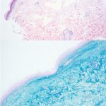
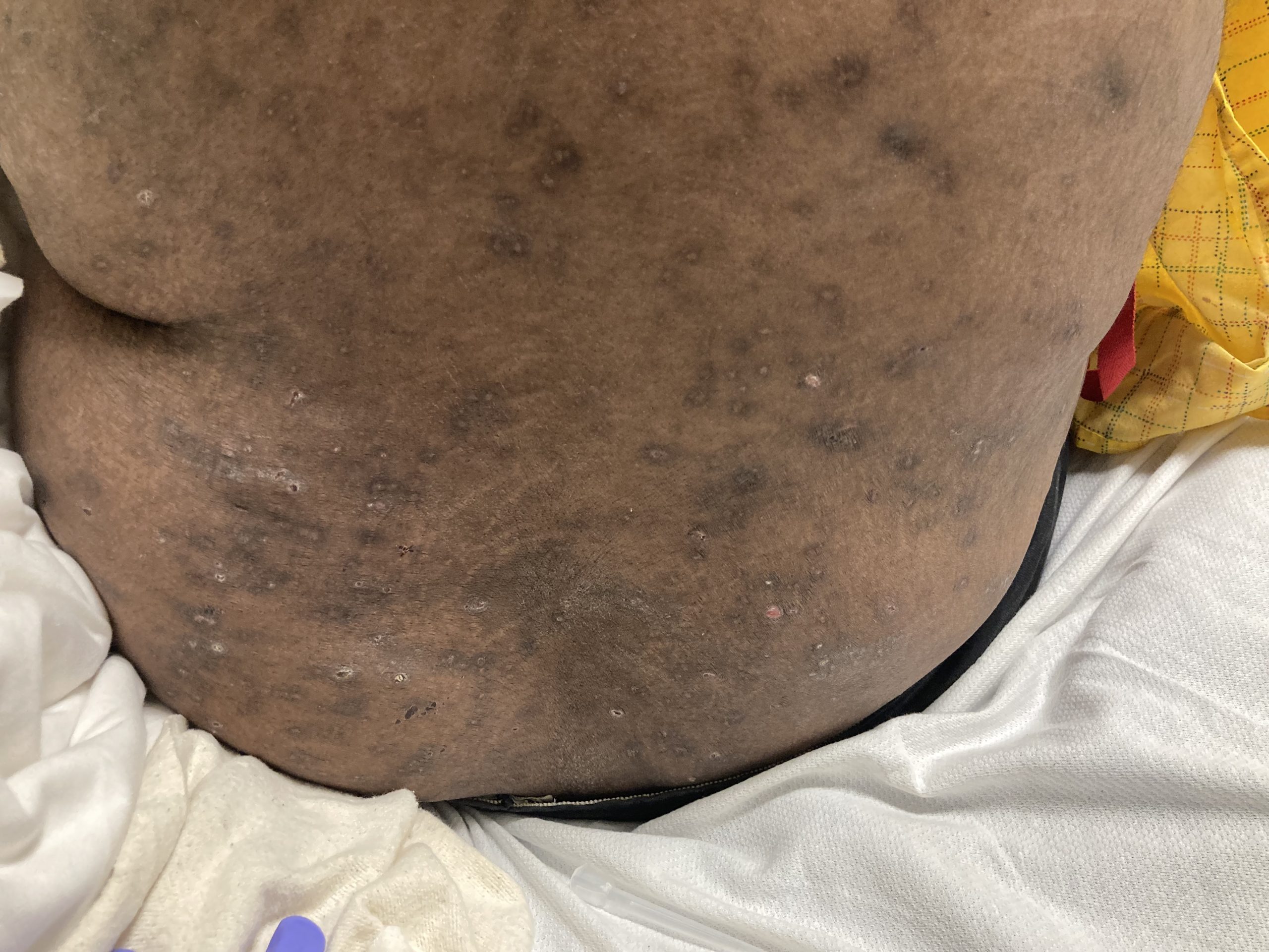
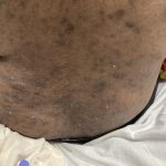
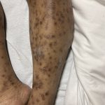
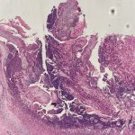
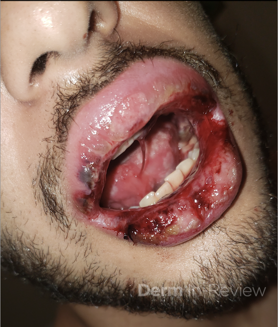
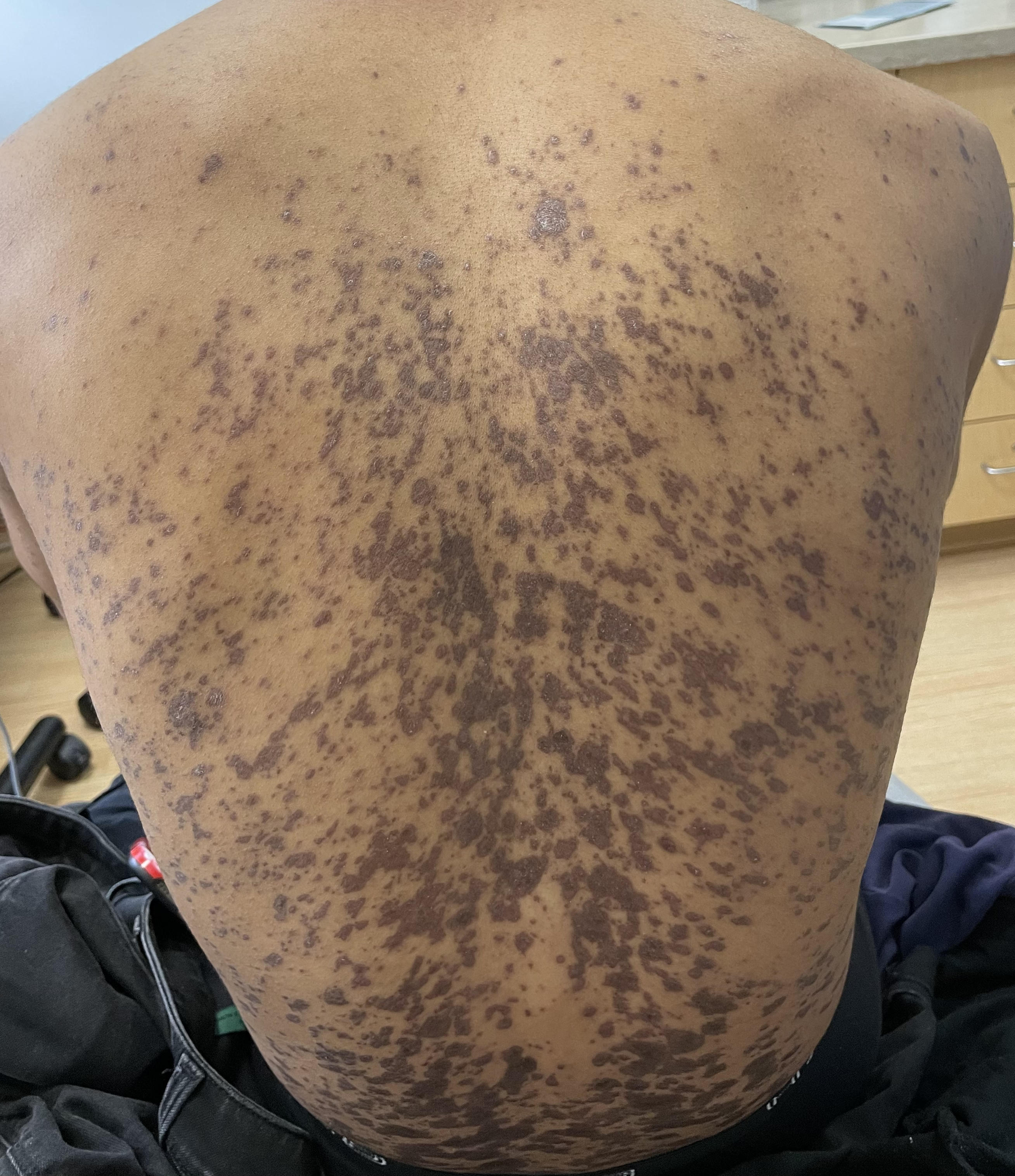
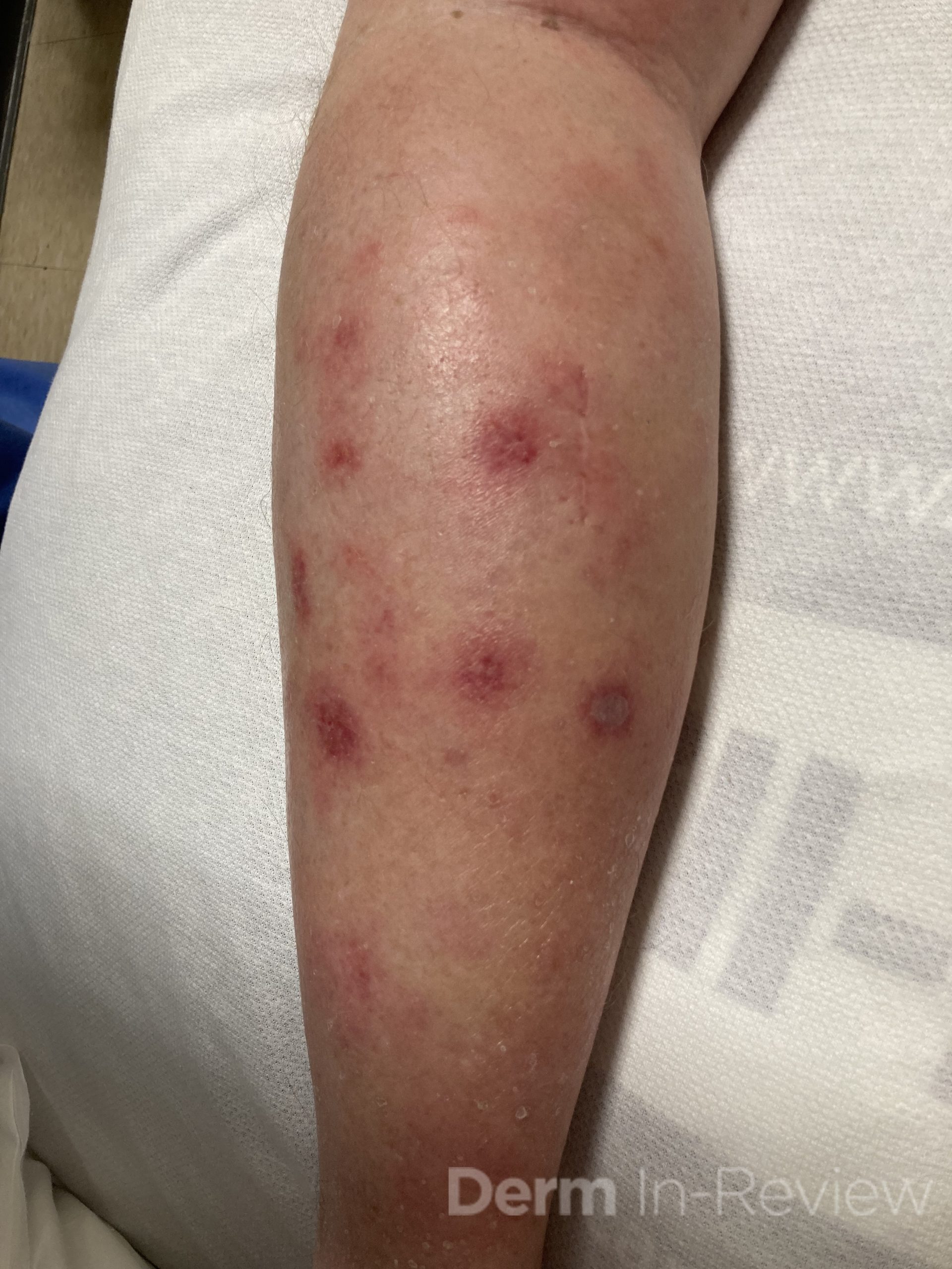
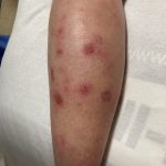
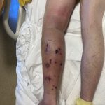
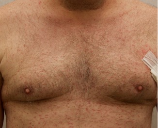
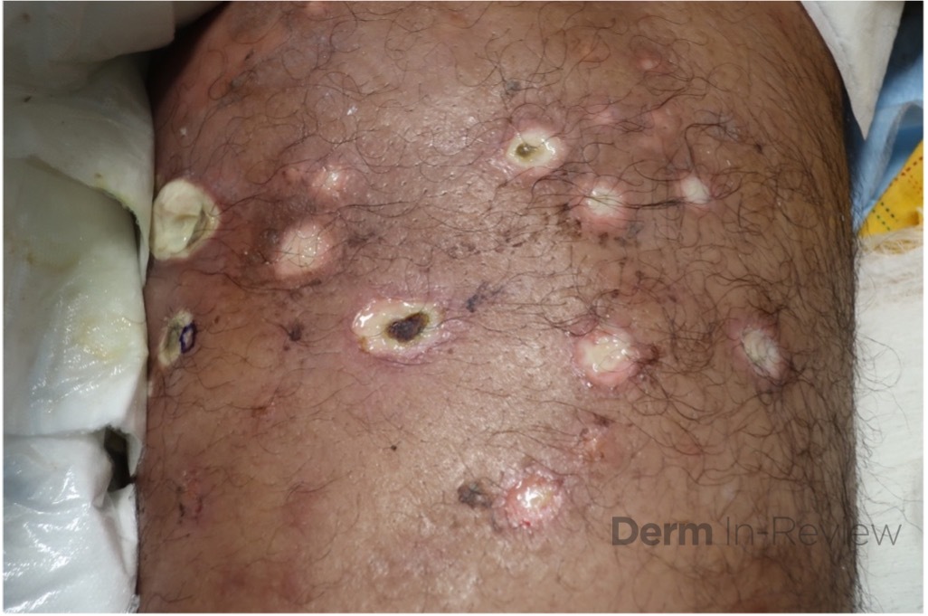
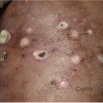
A 43-year-old gentleman with known history of Langerhans cell sarcoma (s/p numerous rounds of chemotherapy and targeted therapy) presented to the ED with a 1-month history of worsening back pain and malaise. He was admitted due to concern for sepsis and started on broad spectrum antibiotics. Dermatology was consulted for painful lesions on his legs that the patient had noticed several months prior to presentation. On examination, the patient had multifocal ulcerations on his bilateral thighs, with overlying fibrinous material (figure 1). A punch biopsy was done on one of these ulcerations (figure 2).  Although these ulcerations could represent several differential diagnoses, the primary concern was whether his known malignancy had metastasized to the skin.

Given his known malignancy history, which stain would be most useful to highlight the underlying pathology?

A.) Factor XIIIA+
B.) CD21+
C.) CD207+
D.) CD31+
E.) S100+

Correct answer: C.) CD207+

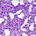
Given the multifocal ulcerations found on exam and pathologic findings of an infiltrate predominantly throughout the dermis and diving deep into the subcutaneous tissue, a malignant process was suspected. The infiltrative cells stained positive for CD207, as well as CD1a and S100. The infiltrate had frequent atypical cells and mitotic figures. As such, the patient was diagnosed with Langerhans cell sarcoma, now with cutaneous metastasis. Langerhans cell sarcoma is a rare neoplastic disorder of Langerhans cells1. According to the Histiocyte Society, there are 5 groups in classifying histiocytoses and neoplasms of the dendritic cell lineages. Langerhans cell sarcoma falls under the malignant, or “M group,” and is a distinct entity from Langerhans cell histiocytosis2. However, Langerhans cell sarcoma will stain positive for typical Langerhans cell markers. These are CD207 (Langerin), CD1a and S100. However, CD207 and CD1a are the most specific stains for Langerhans cells, either of which would have been correct for this question3. Although S100 stain positive for these cells, it also can stain positive for melanocytic proliferations amongst others, so would not be most useful in this case7.

Langerhans cell sarcoma has been diagnosed less than 100 times in the literature1. It can present with both cutaneous and systemic findings, often with multiorgan involvement. There does not appear to be a certain age predilection, as a systematic review in 2015 documented an age range of diagnosis as 11 months to 81 years.1 In this same review, 75% of patients had lymph node involvement, approximately 50% had cutaneous involvement and numerous patients had lung, liver or spleen involvement1. The cutaneous manifestations for this disease process vary from ulcerations to fungating masses, to more indolent appearing nodules. Given its rarity, no consistent presenting cutaneous morphology or lesion is identifiable. The pathogenesis is not well elucidated but is thought to be driven by mutations in the RAS/MAPK/BRAF pathway4. Diagnostic criteria include malignant cytological features such as atypia, hyperchromia and atypia as well as expression of typical Langerhans cell markers as discussed above1. Clear treatment guidelines do not exist, but include surgical excision, radiation, chemotherapy or bone marrow transplantation1. Additionally, targeted therapies against PD-1 and BRAF have shown promise in case reports however no large studies have been published looking at the efficacy of these5,6. The prognosis is poor, particularly in metastatic disease but can be favorable if presenting with one site disease1.

Incorrect stains in answer choices7

Factor XIIIA (choice A) – stains dermal dendritic cells, positive in dermatofibroma and negative in DFSP

CD21 (choice B) – follicular dendritic cell marker, highlights residual follicle in lymphoma

CD31 (choice D) – helpful in confirming vascular origin of tumors, more specific than CD34 for vascular processes

S100 (choice E) – stains melanocytes, Langerhans cells, sweat glands, nerves, Schwann cells, myoepithelial cells, fat, muscle and chondrocytes

References

  1. Langerhans Cell Sarcoma: A Systematic Review.” Cancer Treatment Reviews, U.S. National Library of Medicine
  2. Revised Classification of Histiocytoses and Neoplasms of the Macrophage-Dendritic Cell Lineages.” Blood, U.S. National Library of Medicine
  3. Mizumoto N, Takashima A. CD1a and langerin: acting as more than Langerhans cell markers. J Clin Invest. 2004 Mar;113(5):658-60. doi: 10.1172/JCI21140. PMID: 14991060; PMCID: PMC351325.
  4. Gounder, Mrinal M, et al. “Trametinib in Histiocytic Sarcoma with an Activating MAP2K1 (MEK1) Mutation.” The New England Journal of Medicine, U.S. National Library of Medicine, 17 May 2018,
  5. Zanwar, Saurabh, et al. “Prolonged Remission with Pembrolizumab and Radiation Therapy in a Patient with Multisystem Langerhans Cell Sarcoma.” Haematologica, U.S. National Library of Medicine, 1 Sept. 2022, https://www.ncbi.nlm.nih.gov/pmc/articles/PMC9425309/.
  6. C;Tazi A; “Dramatic Transient Improvement of Metastatic BRAF(V600E)-Mutated Langerhans Cell Sarcoma under Treatment with Dabrafenib.” Blood, U.S. National Library of Medicine, https://pubmed.ncbi.nlm.nih.gov/26468227/.
  7. Elston, D. M., Ferringer, T., & Ko, C. J. (2019).Dermatopathology. Elsevier.

November 2022 Case Study

By 2022 Case Studies

November 2022 Case Study

by Erika McCormick, BS, Emily Murphy, MD, Adam Friedman, MD

A 42-year-old female presented with a rash of two years duration affecting the hands, upper arms, upper back, and chest (Figure 1). A punch biopsy from the back was performed (Figure 2). Laboratory analysis was significant for transcriptional intermediary factor (TIF)-1γ antibody positivity.

Which of the following has been associated with TIF-1γ antibody positivity in this condition?

A.) Mild or absent muscle disease
B.) Increased risk of malignancy
C.) Decreased risk of malignancy
D.) Increased risk of interstitial lung disease
E.) Immune-mediated necrotizing myopathy

Correct answer: B.) Increased risk of malignancy

This patient presented with skin changes consistent with dermatomyositis (DM), including erythematous papules overlying the knuckles (Gottron papules), photodistributed poikiloderma involving the chest (‘V-neck’ sign) and upper back (‘shawl’ sign), and erythema surrounding the periocular region (heliotrope rash; not pictured).2 Punch biopsy from the back showed vacuolar interface dermatitis with necrotic keratinocytes and increased superficial dermal mucin by colloidal iron staining, consistent with DM. A myositis specific antibody panel was performed. Myositis-specific autoantibodies (MSAs) are found in approximately 60 to 70% of children and adults with idiopathic inflammatory myopathy (encompasses DM, inclusion body myositis, polymyositis, immune-mediated necrotizing myopathy, and anti-synthetase syndrome).3 MSAs tend to be mutually exclusive, therefore their detection on laboratory analysis can help identify distinct subsets of disease, predict clinical phenotype, and stratify risk for malignancy and interstitial lung disease.3

In adults greater than 40 years old with DM, anti-TIF1γ autoantibody positivity has a strong association with increased risk of malignancy.3 (Choice B) Anti-nuclear matrix protein 2 (NXP2) is the second most common MSA seen in DM patients with malignancy.3,4 Conversely, antibodies to Mi2 are associated with decreased risk of malignancy.3 (Choice C) Prevalence of malignancy in DM is estimated at 15% to 25%2 with greatest risk within the first 3 years of diagnosis.5 Studies suggest that over 50% of anti-TIF1γ antibody positive DM patients may develop malignancy,6,7and that risk increases to nearly 75% if anti-TIF1α antibodies are also present.6 Conventional recommendations upon diagnosis of DM with elevated malignancy risk include CT scan of the chest, abdomen, and pelvis in addition to obtaining thorough medical history, physical exam, laboratory tests, and age-appropriate cancer screenings.2,8–10 Positron emission tomography with fluorodeoxyglucose and CT (FDG-PET/CT) was found to be comparable to CT alone but may be associated with an increased biopsies without improved diagnostic yield.9,11

DM patients with anti-TIF1 autoantibodies typically have extensive skin involvement but have decreased risk of interstitial lung disease (ILD). 12 (Choice D) Antibodies associated with increased prevalence and severity of ILD include those to aminoacyl-tRNA synthetases (especially PL7, PL12, KS, Jo1, and OJ), PM/Scl, and melanoma differentiation associated protein 5 (MDA-5).3 In anti-MDA-5 antibody positive DM, ILD prevalence ranges from 50% to 100% and can be rapidly progressive with high mortality.13 MDA-5 antibodies are also associated with a characteristic phenotype of mucocutaneous ulcerations, arthritis, and mild or absent muscle disease.3 (Choice A) Immune-mediated necrotizing myopathy, characterized by severe muscle disease refractory to conventional immunosuppressive therapies,3 is associated with antibodies to signal recognition particle (SRP) and to 3-hydroxy-3-methylglutaryl CoA reductase (HMGCR).3 (Choice E)

The treatment approach for DM varies based on severity and extent of myositis; however, it generally includes systemic corticosteroids and immunosuppressive drugs. This patient received a prednisone taper, topical clobetasol 0.05% ointment for rash on the body, and topical hydrocortisone 2.5% ointment for the face. After failing methotrexate, hydroxychloroquine, and mycophenolate mofetil, this patient was successfully transitioned to high dose IVIG. A CT scan of the chest, abdomen, and pelvis was negative for any signs of malignancy and the patient continued age-appropriate cancer screenings with her primary care physician.


References

  1. Wolstencroft PW, Rieger KE, Leatham HW, Fiorentino DF. Clinical factors associated with cutaneous histopathologic findings in dermatomyositis. J Cutan Pathol. 2019;46(6):401-410.
  2. Bolognia J, Schaffer JV, Cerroni L. Dermatology. Fourth edition. (Bolognia J, Schaffer JV, Cerroni L, eds.). Elsevier; 2018.
  3. McHugh NJ, Tansley SL. Autoantibodies in myositis. Nat Rev Rheumatol. 2018;14(5):290-302.
  4. Fiorentino DF, Chung LS, Christopher-Stine L, et al. Most patients with cancer-associated dermatomyositis have antibodies to nuclear matrix protein NXP-2 or transcription intermediary factor 1γ. Arthritis Rheum. 2013;65(11):2954-2962.
  5. Kardes S, Gupta L, Aggarwal R. Cancer and myositis: Who, when, and how to screen. Best Pract Res Clin Rheumatol. 2022;36(2).
  6. Fujimoto M, Hamaguchi Y, Kaji K, et al. Myositis-specific anti-155/140 autoantibodies target transcription intermediary factor 1 family proteins. Arthritis Rheum. 2012;64(2):513-522.
  7. Hoshino K, Muro Y, Sugiura K, Tomita Y, Nakashima R, Mimori T. Anti-MDA5 and anti-TIF1-gamma antibodies have clinical significance for patients with dermatomyositis. Rheumatology (Oxford). 2010;49(9):1726-1733.
  8. Oldroyd AGS, Allard AB, Callen JP, et al. A systematic review and meta-analysis to inform cancer screening guidelines in idiopathic inflammatory myopathies. Rheumatology (Oxford). 2021;60(6):2615-2628.
  9. Maliha PG, Hudson M, Abikhzer G, Singerman J, Probst S. 18F-FDG PET/CT versus conventional investigations for cancer screening in autoimmune inflammatory myopathy in the era of novel myopathy classifications. Nucl Med Commun. 2019;40(4):377-382.
  10. Vaughan H, Rugo HS, Haemel A. Risk-Based Screening for Cancer in Patients With Dermatomyositis: Toward a More Individualized Approach. JAMA Dermatol. 2022;158(3):244-247.
  11. Selva-O’Callaghan A, Grau JM, Gámez-Cenzano C, et al. Conventional cancer screening versus PET/CT in dermatomyositis/polymyositis. Am J Med. 2010;123(6):558-562.
  12. Fiorentino DF, Kuo K, Chung L, Zaba L, Li S, Casciola-Rosen L. Distinctive cutaneous and systemic features associated with antitranscriptional intermediary factor-1γ antibodies in adults with dermatomyositis. J Am Acad Dermatol. 2015;72(3):449-455.
  13. Wu W, Guo L, Fu Y, et al. Interstitial Lung Disease in Anti-MDA5 Positive Dermatomyositis. Clin Rev Allergy Immunol. 2021;60(2):293-304.

October 2022 Case Study

By 2022 Case Studies

October 2022 Case Study

by Emily Murphy, MD

A 36-year-old male with a past medical history of HIV/AIDS (CD4 count of 3) presented with a diffuse pruritic rash of several weeks duration. The rash started on his arms and then spread to his legs, back, and chest. On examination, the patient had innumerable papules of varying morphologies, including many lichenified papules with keratotic cores (figure 1). A punch biopsy was done of a papule on the leg (figure 2).

Based on the clinical presentation and biopsy findings, which stain would be most useful to highlight the underlying pathology?

A.) Toluidine blue
B.) Verhoeff–Van Gieson stain
C.) Colloidal iron
D.) Congo red
E.) Von Kossa

Correct answer: B.) Verhoeff–Van Gieson stain

Given the papules with central keratotic cores present on exam and pathologic finding of a crateriform erosion with collagen extrusion, the patient was diagnosed with acquired perforating dermatosis in the setting of HIV. The clinical findings are due to the transepidermal elimination of collagen through the epidermis; the extruded collagen can be stained red with the Verhoeff–Van Gieson stain (choice B). This entity is classically seen in the setting of chronic kidney disease, often requiring dialysis, and diabetes mellitus, most often with associated nephropathy.1 Acquired perforating dermatosis can also be seen due to numerous medications (TNFα inhibitors, epidermal growth factor inhibitors, sirolimus, or antivirals like indinavir)1,2 and has been reported in the setting of HIV.2–4

Acquired perforating dermatosis most commonly occurs on the legs but can be generalized, as in our patient. The head is typically minimally involved and it spares the palms/soles, intertriginous areas, and mucous membranes.4  Papules and nodules are seen, with the biggest clue to the diagnosis being the central keratotic cores.1 The eruption is very pruritic; scratching can trigger further disease via the Koebner phenomenon, and the microtrauma induced by scratching may also trigger focal degeneration of collagen, contributing to the pathogenesis.2 In addition to skin trauma, increased growth factors, such as TGF-β, and fibroblast activity may also play a role in the disease.2 On histopathology, a well-circumscribed, cup-shaped depression bordered by epidermal hyperplasia is seen with vertically-oriented, degenerated, basophilic collagen fibers extruding through the epidermis.2,5

Management includes treatment of the underlying disease trigger and symptomatic treatment of pruritus, using topical steroids, antipruritic emollients, keratolytics (salicylic acid, urea), or oral antihistamines.5 Phototherapy (typically NB-UVB) can also treat pruritus and reduce skin disease.5 The lesions often heal with post-inflammatory pigmentary changes or atrophic scars.2,5

Incorrect stains in answer choices:6

Toluidine blue (choice A) – mast cells and mucin

Colloidal iron (choice C) – acid mucopolysaccharides (mucin)

Congo red (choice D) – amyloid

Von Kossa (choice E) – calcium salts


References

  1. Rapini RP. Chapter 96 – Perforating Diseases. In: Dermatology. Fourth. Elsevier.
  2. Wagner G, Sachse MM. Acquired reactive perforating dermatosis. J Dtsch Dermatol Ges. 2013;11(8):723-729, 723-730. doi:10.1111/ddg.12131
  3. Rubio FA, Herranz P, Robayna G, Peña JM, Contreras F, Casado M. Perforating folliculitis: report of a case in an HIV-infected man. J Am Acad Dermatol. 1999;40(2 Pt 2):300-302. doi:10.1016/s0190-9622(99)70470-6
  4. Bank DE, Cohen PR, Kohn SR. Reactive perforating collagenosis in a setting of double disaster: acquired immunodeficiency syndrome and end-stage renal disease. J Am Acad Dermatol. 1989;21(2 Pt 2):371-374. doi:10.1016/s0190-9622(89)80037-4
  5. Lukács J, Schliemann S, Elsner P. Treatment of acquired reactive perforating dermatosis – a systematic review. J Dtsch Dermatol Ges. 2018;16(7):825-842. doi:10.1111/ddg.13561
  6. Ferringer T, Ko CJ. Chapter 1 – The basics. In: Dermatopathology. Third. Elsevier.

September 2022 Case Study

By 2022 Case Studies

September 2022 Case Study

by Alexis Carrington, MD


A 22 year-old male with a past medical history of asthma presents to the ED with a rash on the mouth and bilateral palms that appeared a day prior to presentation. A week prior, he went to the ED for worsening throat and mouth pain, where he was diagnosed with thrush and given nystatin but didn’t have any skin manifestations at that time. He also reported subjective fevers, chills, decreased appetite, fatigue and non-bloody diarrhea that started two days ago. Exam showed hemorrhagic mucositis with some minor re-epithelialization and a papular target lesions on the hands and left arm. Based on examination, a diagnosis of Erythema Multiforme was made.

What is the most common precipitating factor for Erythema Multiforme?

A.) Herpes Simplex Virus (HSV)
B.) Parapoxvirus
C.) Mycoplasma pneumoniae
D.) Histoplasma capsulatum


Correct answer: A.) Herpes Simplex Virus (HSV)

Erythema Multiforme (EM) is an immune-mediated mucocutaneous condition characterized by “target” lesions. Severe mucosal involvement is what distinguishes erythema multiforme major from erythema multiforme minor. EM episodes can be recurrent or persistent, but are typically self-limiting. Some triggers of EM include infections, as well as medications and even vaccinations.

The characteristic morphology of EM are typical target lesions, which are a round shape with a well defined border and consist of three distinct zones distributed on the extremities and the face, the dorsal hands and forearms being the most frequently involved. Mucosal lesions of EM are vesiculobullous and develop into painful erosions on the buccal mucosa and lips. Diagnosis is clinical but not based solely on histological findings.

The most common associated infectious agent with EM is Herpes Simplex Virus (answer A). Parapoxvirus (answer B) Histoplasma capsulatum (answer D) and Mycoplasma pneumoniae (answer C) are observed less frequently.

Treatment is supportive as EM episodes are typically self limiting. However, early therapy with systemic corticosteroids can be considered in severe forms of EM with functional impairment.


References

  1. Canavan TN, Mathes EF, Frieden I, Shinkai K. Mycoplasma pneumoniae-induced rash and mucositis as a syndrome distinct from Stevens-Johnson syndrome and erythema multiforme: a systematic review. J Am Acad Dermatol. 2015;72(2):239–45. doi:10.1016/j.jaad.2014.06.026.
  2. Soares A, Sokumbi O. Recent Updates in the Treatment of Erythema Multiforme. Medicina (Kaunas). 2021;57(9):921. Published 2021 Sep 1. doi:10.3390/medicina57090921.
  3. Bolognia, Jean L. Dermatology volume 2. Mosby, 2018.

August 2022 Case Study

By 2022 Case Studies

August 2022 Case Study

by Blair Allais, MD

A 55-year-old male presented to clinic with a new worsening red, scaly rash of the forehead, temples and upper cheeks. He had tried to use over the counter hydrocortisone and witch hazel toner at home without improvement of the rash. On further questioning of possible exposures, the patient endorsed switching his home hair dye recently and used it for the second time several days prior to the development of the rash.

Which of the following cross reacts with the likely culprit allergen in this case?

A.) Aspirin
B.) Penicillin
C.) Glipizide
D.) Lidocaine
E.) Furosemide

Correct answer: C.) Glipizide

Para-phenylenediamine (PPD) dye is commonly found in many consumer products including hair dye, black henna tattoos, textiles, leather, fur, printer ink, and black rubber products.1 The majority of reactions to topical PPD are delayed-type IV hypersensitivity in nature and characterized by pruritus, erythema, and vesiculobullous eruptions that may occur on the face, scalp margins, or ears.2 Cross-sensitization to other compounds that also contain a benzene ring at the para position can occur.3 A helpful mnemonic to remember the chemicals that cross-react with PPD can be found in Chapter 2 of the Derm-In-Review Text: “AA SSTEPP”, which stands for Azo, Aniline dyes, Sulfonamides, Sulfonylurea, Thiazides, Esters (benzocaine), Procainamide, PABA and PABA esters.


Glipizide is a sulfonylurea and cross reacts with PPD. The other answer choices are distractors and incorrect. Para-aminosalicylic acid (an anti-tuberculosis medication) is a cross reactor to PPD dye, and one should not confuse para-aminosalicylic acid with aspirin (acetyl salicylic acid). While sulfonamides cross react with PPD, other antibiotics such as penicillin do not. While ester anesthetics (procaine, chlorprocaine and tetracaine) cross react with PPD, amide antibiotics such as lidocaine do not.  While thiazide diuretics (hydrochlorothiazide, chlorthalidone) cross react with PPD, loop diuretics such as furosemide do not.


References

  1. Rietschel RL, Fowler JF. Fishers Contact Dermatitis, 4th edn. Baltimore, MD: Williams and Wilkins, 1995.
  2. Mukkanna KS, Stone NM, Ingram JR. Para-phenylenediamine allergy: current perspectives on diagnosis and management. J Asthma Allergy. 2017;10:9-15. Published 2017 Jan 18. doi:10.2147/JAA.S90265
  3. McFadden JP, Yeo L, White JL. Clinical and experimental aspects of allergic contact dermatitis to para-phenylenediamine. Clin Dermatol. 2011;29(3):316-324. doi:10.1016/j.clindermatol.2010.11.011

July 2022 Case Study

By 2022 Case Studies

July 2022 Case Study


A 70-year-old woman with history of hypothyroidism and asthma presents to clinic with a 3-month history of sores in her mouth. She denies any other symptoms, but she does report a 5-pound weight loss due to pain with eating. On further exam, there are erosions of the gingiva, buccal mucosa, and posterior neck. There is no involvement of other mucosal sites. Patient undergoes biopsies for hematoxylin and eosin staining and direct immunofluorescence (DIF). Results showed a subepidermal bullae with positive IgG and C3 staining at the dermo-epidermal junction. Further testing revealed presence of anti-laminin 332 antibodies.

What additional work up is needed?

A.) Referral to ophthalmology
B.) Pulmonary function testing
C.) Perform all age-appropriate cancer screenings
D.) Endoscopy
E.) No additional work up needed

Correct answer: C.) Perform all age-appropriate cancer screenings

Mucous membrane pemphigoid (MMP) is a subepidermal autoimmune blistering disorder with various phenotypes. As the name suggests, clinical features involve erosions of mucosal surfaces, often with subsequent scarring. The most commonly affected mucosal sites are oral and ocular surfaces. Those with ocular involvement are at risk for blindness so it is important to ensure ophthalmologic monitoring when necessary. Non-mucosal skin can be affected, but this occurs in less than 50% of patients.1


Previously, subtypes were classified based on antigenic targets or area of involvement – anti-elipigrin cicatricial MMP (laminin 332), ocular MMP (b4 subunit of integrin a6b4), anti-bullous pemphigoid antigen MMP (BPAG180 or BPAG230).1 Additionally, the “Brunsting-Perry” variant classified a subset of patients with cutaneous involvement of the head and neck resulting in a cicatricial alopecia.1 However, recent updates in nomenclature have moved away from use of these subtypes and toward use of phenotypic descriptions.2


Diagnosis of MMP requires a combined approach as diagnosis can often be delayed. As with other vesiculobullous diseases, DIF is essential to diagnosis and has been found to be the most sensitive diagnostic tool.3,4 Classic findings on DIF are IgG, IgA, and/or C3 deposition at the dermo-epidermal junction.4 Salt split skin technique is useful to differentiate likely antigenic targets. Roof staining with IgG or IgA is consistent with BP180 or BP230 targets while floor staining with IgG or IgA correlates to laminin 332 or type VII collagen targets.4 It is important to remember that anti-laminin 332 MMP, as seen in this patient, is associated with an increased risk of malignancy.1,4  Thus, it is important that patients with this variant undergo age-appropriate cancer screenings at the time of diagnosis.


References

  1. Bolognia JK, Schaffer JV, Cerroni L. Dermatology. Philadelphia: Elsevier. 2018.
  2. Rashid H, et al. European guidelines (S3) on diagnosis and management of mucous membrane pemphigoid, initiated by the European Acadmy of Dermatology and Venereology – Part I. 2021;35:1750-1764.
  3. Rashid H et al. Assessment of diagnostic strategy of mucous membrane pemphigoid. JAMA Derm. July 2021;157(7):780-787.
  4. Schmidt E, et al. European guidelines (S3) on diagnosis and management of mucous membrane pemphigoid, initiated by the European Academy of Dermatology and Venereology – Part II. JEADV. 2021;35:1926-1948.

June 2022 Case Study

By 2022 Case Studies

June 2022 Case Study

by Adrianna Gonzalez, MD

A 29 year old male with no significant past medical history presented with complaints of a generalized rash which began two months prior. He stated the eruption began as a few lesions in the trunk that progressively grew in number and generalized to involve upper and lower extremities, buttocks, and genitals. He stated rash was associated with significant itch. Of note, he stated he had taken a new medication several weeks prior to the onset of his rash. Findings on clinical examination can be seen in Figure 1. A 4mm punch biopsy of a lesion on his mid back was performed. Histopathological examination revealed orthokeratosis, hypergranulosis, acanthosis with sawtoothing rete ridge pattern, dyskeratotic keratinocytes and a band-like lymphocytic infiltrate.

Which of the following statements is FALSE regarding this patient’s condition?

A.) The oral mucosa is the most commonly involved body site in this entity
B.) On histology, dyskeratotic keratinocytes are classically found at the upper level of the epidermis
C.) Direct immunofluorescence may be positive for “shaggy” fibrinogen deposits along the dermoepidermal junction
D.) Flares may be triggered by certain contact allergens including copper
E.) The majority of cases will remit within 2 years

Correct answer: B.) On histology, dyskeratotic keratinocytes are classically found at the upper level of the epidermis

The clinical and histopathological findings in this case are consistent with lichen planus (LP). On histology, LP is characterized by compact orthokeratosis, wedge-shaped hypergranulosis, sawtooth pattern of rete ridges, vacuolar degeneration of the basal layer, dyskeratotic keratinocytes (Civatte bodies), and a band-like lymphocytic infiltrate at the dermoepidermal junction (DEJ). Dyskeratotic keratinocytes are classically present at the lower levels of the epidermis and at the superficial papillary dermis near the basal layer, the site of destruction.1 Dyskeratotic keratinocytes can be seen in the suprabasilar, upper epidermal layer in erythema multiforme, Stevens-Johnson Syndrome and toxic epidermal necrolysis.2 Direct immunofluorescence (DIF) demonstrates “shaggy” deposition of fibrinogen along the basement membrane zone, as well as staining of colloid bodies with one or more of IgM, IgG, IgA and C3  (answer C).


Lichen planus is an inflammatory disorder of the skin, mucosa, hair and nails. Although the incidence appears to vary throughout geographical areas, the oral mucosa has been found to be the most commonly involved body site in most study populations (answer A).1,3 Oral lesions can be observed in over 50% of patients with LP, and can often be the only site of involvement.2 A recent epidemiological study estimated that the global pooled prevalence of oral LP was 0.89% among the general population and 0.98% among clinical patients.4 Cutaneous LP is thought to affect < 1% of the population worldwide and commonly presents as pruritic, purple, polygonal, flat-topped papules commonly involving the extremities and lower back.5 There are numerous variants of cutaneous LP, including actinic, annular, atrophic, bullous, drug-induced, genital, hypertrophic, inverse, linear, LP pigmentosus and lichen planopilaris. LP may also involve the nails and present with longitudinal ridging, trachyonychia, thinning, dorsal pterygium and even 20 nail dystrophy. While its pathophysiology has not been fully elucidated, LP is thought to be caused by an altered cellular immune response, in which cytotoxic CD8+ T cells attack basal keratinocytes. LP has been associated with various triggers, including infections such as hepatitis C virus (more common in oral erosive LP), medications, contact allergens, vaccinations (hepatitis B virus and influenza) and genetics. Implicated contact allergens include gold, mercury amalgam and copper (answer D).1  Most cases of LP self-resolve within 1-2 years (answer E). Oral LP (especially oral erosive LP), nail LP and hypertrophic LP tend to be persistent and recalcitrant to treatment.

References

  1. Boch K, Langan EA, Kridin K, Zillikens D, Ludwig RJ, Bieber K. Lichen Planus. Front Med (Lausanne). 2021;8:737813.
  2. Bolognia J, Jorizzo, J. L., & Schaffer, J. Dermatology. Fourth Edition ed. Philadelphia: Elsevier Saunders; 2012.
  3. Arnold DL, Krishnamurthy K. Lichen Planus. In: StatPearls. Treasure Island (FL)2022.
  4. Li C, Tang X, Zheng X, et al. Global Prevalence and Incidence Estimates of Oral Lichen Planus: A Systematic Review and Meta-analysis. JAMA Dermatol. 2020;156(2):172-181.
  5. Tziotzios C, Lee JYW, Brier T, et al. Lichen planus and lichenoid dermatoses: Clinical overview and molecular basis. J Am Acad Dermatol. 2018;79(5):789-804.

May 2022 Case Study

By 2022 Case Studies

May 2022 Case Study

by Emily Murphy, MD

A 65-year-old male with a past medical history of HIV (CD4 count 250, undetectable viral load) and chronic congestion presented with a chief complaint of recurrent cellulitis of the left lower leg. The redness and swelling persisted for 3 months despite antibiotics. On presentation, he had circumferential edema and induration of the left lower leg with erythematous, tender nodules and early bullae (figure 1). A week later, his disease progressed with ulceration and necrosis of these tender nodules (figure 2). A punch biopsy was done, which showed an atypical lymphoid infiltrate in the dermis and subcutaneous tissue with an angiocentric pattern, composed of large, pleomorphic cells. Extensive necrosis was present with karyorrhetic debris. Immunostains were positive for T cells (CD2), natural killer cells (CD56), and cytotoxicity (granzyme B). A biopsy of the nasal septum showed similar findings. The patient was diagnosed with extranodal NK/T cell lymphoma, nasal type.

Which virus is part of the diagnostic criteria for this lymphoma?

A.) Human herpesvirus-8
B.) Polyomavirus
C.) Human immunodeficiency virus
D.) Ebstein Bar Virus
E.) Cytomegalovirus

Correct answer: D.) Ebstein Bar Viru

Extranodal NK/T cell lymphoma, nasal type is an aggressive, non-Hodgkin lymphoma that has four criteria according to the World Health Organization: vascular damage, prominent necrosis, a cytotoxic phenotype, and association with the Ebstein bar virus (EBV, answer D).1,2 EBV positivity is seen in the both the skin and serum. Pathologically, the atypical lymphocytes are positive for cytotoxic markers (granzyme B, peforin), natural killer cell markers (CD2), T cell markers (cytoplasmic CD3), and EBV.1 Extranodal NK/T cell lymphoma is rare in the United States, but its incidence is increasing. It is more common in East Asia and Latin America.1

This lymphoma typically affects the upper aerodigestive tract, causing congestion, epistaxis, or necrotizing lesions of the nose or hard palate.1 Other sites can be involved, including the extranasal skin, gastrointestinal tract, testes, or lungs.1,3,4 On the skin, either a generalized morbilliform eruption or purpuric nodules with or without ulceration can be seen.3 Hemophagocytic lymphohistiocytosis can occur, but is rare, occurring in only 3% of patients.1

NK/T cell lymphoma has an increased incidence in patients with HIV; a study of 93 patients with this lymphoma found that it occurred at a 15 times higher incidence in patients with HIV compared to the general population.5 However, HIV (choice C) is not part of the diagnostic criteria like EBV. Human herpesvirus-8 (Kaposi sarcoma), Polyomavirus (Merkel Cell Carcinoma), and cytomegalovirus are not associated with Extranodal NK/T cell lymphoma (choices A, B, E).

Treatment is determined by the Ann Arbor Staging and various prognostic tools, like the PINK-E system (age > 60 years, Stage III/IV, nonnasal primary localization, distant lymph node involvement, and detectable plasma EBV DNA).6 Depending on the stage and prognosis, treatment is typically with an asparaginase-based regimen called SMILE (dexamethasone, methotrexate, ifosfamide, L-asparaginase, and etoposide).1,6,7 NK/T cell lymphoma cells are unable to synthesize the amino acid, asparagine, and as this amino acid is depleted with asparaginase, protein synthesis is disrupted and the cells ultimately undergo apoptosis.6 Chemotherapy is typically followed by involved field radiation therapy.1  Various immunotherapies are currently being studied for patients with recalcitrant or relapsed disease as well, including programmed death 1 inhibitors, JAK 3 inhibitors, or anti-CD30 therapies.8 Response to therapy can be monitored with EBV DNA titers, which should decrease with treatment.1

References

  1. Haverkos BM, Pan Z, Gru AA, et al. Extranodal NK/T Cell Lymphoma, Nasal Type (ENKTL-NT): An Update on Epidemiology, Clinical Presentation, and Natural History in North American and European Cases. Curr Hematol Malig Rep. 2016;11(6):514-527. doi:10.1007/s11899-016-0355-9
  2. Willemze R, Cerroni L, Kempf W, et al. The 2018 update of the WHO-EORTC classification for primary cutaneous lymphomas. Blood. 2019;133(16):1703-1714. doi:10.1182/blood-2018-11-881268
  3. Orlowski GM, Tan AJ, Evan-Browning E, Scharf MJ. Primary cutaneous nasal-type NK/T-cell lymphoma presenting as purpuric nodules on the lower leg. JAAD Case Rep. 2020;6(10):1075-1078. doi:10.1016/j.jdcr.2020.08.007
  4. Rahal A, Reddy PS, Alvares C. Extranodal NK/T-Cell Lymphoma, Nasal Type, Presenting as a Breast Mass. Cureus. 7(12):e408. doi:10.7759/cureus.408
  5. Castillo J, Pantanowitz L. HIV-Associated NK/T-Cell Lymphomas: A Review of 93 Cases. Blood. 2007;110(11):3457-3457. doi:10.1182/blood.V110.11.3457.3457
  6. van Doesum JA, Niezink AGH, Huls GA, Beijert M, Diepstra A, van Meerten T. Extranodal Natural Killer/T-cell Lymphoma, Nasal Type: Diagnosis and Treatment. Hemasphere. 2021;5(2):e523. doi:10.1097/HS9.0000000000000523
  7. Yamaguchi M, Kwong YL, Kim WS, et al. Phase II study of SMILE chemotherapy for newly diagnosed stage IV, relapsed, or refractory extranodal natural killer (NK)/T-cell lymphoma, nasal type: the NK-Cell Tumor Study Group study. J Clin Oncol. 2011;29(33):4410-4416. doi:10.1200/JCO.2011.35.6287
  8. Lv K, Li X, Yu H, Chen X, Zhang M, Wu X. Selection of new immunotherapy targets for NK/T cell lymphoma. Am J Transl Res. 2020;12(11):7034-7047.

April 2022 Case Study

By 2022 Case Studies

April 2022 Case Study

by Blair Allais, MD

A 63-year-old male presented to clinic with a 2-year history of asymptomatic annular hypopigmented plaques on the trunk and extremities that eventually became erythematous to violaceous. He had been prescribed topical steroids by an outside practitioner without improvement in the lesions. Biopsy performed on one of the plaques revealed a granulomatous reaction surrounding adnexal structures and in the papillary dermis. Fite stain revealed numerous pink to red acid-fast organisms within the granulomas ultimately rendering a diagnosis of lepromatous leprosy. The patient was started on therapy with Rifampin 600mg monthly, Minocycline 100mg monthly and Moxifloxacin 400mg monthly. Approximately 3 months after initiating treatment the patient developed new nodular skin lesions on the extensor arms and medial thighs associated with fever, myalgias, fatigue and joint pain.

What is the best course of treatment for this patient’s new presentation?

A.) Prednisone
B.) Clofazimine
C.) Thalidomide
D.) Methotrexate

Correct answer: C) Thalidomide

The patient described has a diagnosis of lepromatous leprosy and is experiencing a reactional state to treatment. Approximately 50% of patients with leprosy will experience a reaction after the institution of therapy. Aside from initiating antibiotic therapy, other causes of reactional states include intercurrent infections, vaccination, pregnancy, vitamin A, iodides and bromide. Reactions are an important cause of permanent nerve damage in borderline patients and are abrupt in appearance.


Two major types of reactional states have been described. Type 1 reactions due to enhancement of cell-mediated immunity with a Th1 cytokine pattern (IFN-y, TNF, IL-1ß, IL-2, IL-12). These reactions are considered to reversal reactions with “upgrading” and are seen predominantly in borderline or tuberculoid forms of leprosy. They are characterized by increased inflammation within existing skin lesions in addition to acute nerve pain or tenderness (neuritis) and loss of function. Treatment of choice for these reactions is prednisone (A) starting at a dose of 40 – 60 mg/day. Neuritis and eye lesions are urgent indications for therapy and dose and duration is determined by the clinical course of the reaction. Steroids are tapered slowly over months to years once the reaction is controlled. In some cases, clofazimine (B) may be added as it has some activity against the type 1 reaction, and may be added in doses of up to 300 mg/day as tolerated.


Type 2 reactions are due to excessive humoral immunity with a Th2 cytokine pattern (IL-4, IL-10) and characterized by formation of immune complexes. They are seen predominately in patients with lepromatous and borderline lepromatous forms of leprosy, especially in patients with a high bacterial load who are undergoing treatment. Multisystem involvement is seen with systemic symptoms including nodular skin lesions, fever, myalgias, malaise, severe joint swelling and pain, iridocyclitis, lymphadenitis, hepatosplenomegaly, orchitis and glomerulonephritis. The intensity of the reaction can vary from mild to severe and can last from a few days to weeks, months, or even years. Pathology is characterized by cutaneous and systemic small vessel vasculitis, and treatment of choice is Thalidomide (C). According to the newly developed U.S. regimen for treatment for lepromatous leprosy, methotrexate (D) 15mg weekly can be prescribed to prevent reactions at the time of initiating multi drug treatment.

References

  1. Ramos-e-Silva M, Ribeiro de Castro MC. Chapter 75: Mycobacterial Infections. 4th Ed. Philadelphia, PA: Elsevier; 2018.
  2. James WD et al. Chapter 17: Hansen Disease. Andrews’ Diseases of the Skin. 13th Philadelphia, PA: Elsevier; 2020.
  3. Bennett JE et al. Principles and Practice of Infectious Diseases. 6th edit.P.2887-2896

March 2022 Case Study

By 2022 Case Studies

March 2022 Case Study

by Daniel Nussbaum, MD

A 50-year-old man with no past medical history presents with the pictured rash on his chest, arms, abdomen, and back for five years. Although asymptomatic, the patient reported increasing redness under hot water without associated pruritus. He denied any systemic symptoms. Examination showed monomorphic erythematous macules and papules distributed diffusely over the chest, upper extremities, abdomen, and back. He denies any systemic symptoms and has no personal or family history of skin conditions other than the pictured lesions.

Which of the following laboratory tests would aid in confirming the diagnosis in this patient?

A.) Indirect immunofluorescence
B.) Anti-gliadin Ab
C.) Anti-nuclear Ab
D.) Anti-Jo-1 Ab
E.) Serum tryptase

Correct answer: E

The history and image provided are most consistent with a adult onset cutaneous mastocytosis. Histopathology of cutaneous mastocytosis commonly reveals mast cells concentrated in the dermal papillae, which can be confirmed with staining for mast cells (see below).1 The patient was recommended to avoid triggers for mast cell degranulation such as heat, sunlight, stress, medications, and alcohol. Additionally, he was referred to hematology for bone marrow biopsy, which was deferred due to lack of systemic symptoms.

Mastocytosis is a spectrum of diseases with various cutaneous and systemic manifestations. Cutaneous findings include flushing, pruritus, urticaria, and bullae. Systemic symptoms include bone pain, headaches, fatigue, nausea, vomitus, diarrhea, weight loss, cramping, chest pain, palpitations, and syncope.1 Patients with mastocytosis, especially the systemic variant, frequently have a mutation in KIT D816V, a tyrosine kinase receptor. Tissue diagnoses of mastocytosis can be confirmed with Giemsa, CD117, Leder, toluidine blue, and tryptase stains.

Diffuse cutaneous mastocytosis is a rare subset of mastocytosis, usually presenting in childhood low risk of systemic involvement. However, when presentation occurs in adulthood, systemic involvement should be evaluated with serum tryptase and bone marrow biopsy when indicated. Without a bone marrow biopsy, the risk of systemic involvement can be estimated from symptoms and a tryptase level.2 Based on clinical presentation and laboratory findings, physicians must consider working up systemic involvement to avoid morbidity and mortality.3,4

Indirect immunofluorescence (IIF) (answer A) is useful in the diagnosis of blistering dermatoses. Specifically, IIF of serum Ab to monkey esophagus can be useful in the diagnosis of pemphigus vulgaris.5 Anti-gliadin Ab (answer B) are commonly found in patients with dermatitis herpetiformis, which is a skin manifestation of gluten-sensitive enteropathy, commonly known as celiac disease.6 Anti-nuclear Ab (answer C) are non-specific for various autoimmune conditions such as systemic lupus erythematosus, rheumatoid arthritis, systemic sclerosis, among others.7 Anti-Jo-1 Ab (answer D) are more specific autoimmune markers for patients with dermatomyositis and polymyositis.8 [/vc_column_text]

References

  1. Bolognia JK, Schaffer JV, Cerroni L. Dermatology. Philadelphia: Elsevier. 2018.
  2. Fuchs D, Kilbertus A, Kofler K, et al. Scoring the risk of having systemic mastocytosis in adult patienst with mastocytosis in the skin. J Allergy Clin Immunol Pract. April 2021;9(4):1705-1712.
  3. Mannelli F. Catching the clinical and biological diversity for an appropriate therapeutic approach in systemic mastocytosis. Ann Hematol. 2021;100:337-344.
  4. Pardanani, A. Systemic mastocytosis in adults: 2021 update on diagnosis, risk stratification and management. Am J Hematol. 2021;96:508-525.
  5. Kridin K, Bergman R. The Usefulness of Indirect Immunofluorescence in Pemphigus and the Natural History of Patients With Initial False-Positive Results: A Retrospective Cohort Study. Front Med (Lausanne). 2018;5:266.
  6. Benson BC, Mulder CJ, Laczek JT. Anti-gliadin antibodies identify celiac patients overlooked by tissue transglutaminase antibodies. Hawaii J Med Public Health. 2013;72(9 Suppl 4):14-17.
  7. Heffernan MP, Do JH, Mehta J. Antinuclear antibodies in dermatology. Semin Cutan Med Surg. 2001;20(1):2-13.
  8. Mileti LM, Strek ME, Niewold TB, Curran JJ, Sweiss NJ. Clinical characteristics of patients with anti-Jo-1 antibodies: a single center experience. J Clin Rheumatol. 2009;15(5):254-255.