February 2020 Case Study
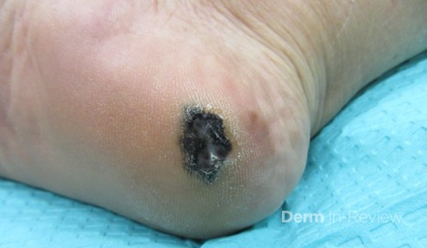
77 year old woman with no personal or family history of skin cancer presented with a lesion on her R heel that she noticed 2 months prior. She had not noticed any growth of the lesion and it was not tender. On physical exam she had a 1.2 x 0.9 cm dark brown/black plaque with hyperkeratosis on the R plantar heel with no lymphadenopathy (Image 1). On pathology, there was an asymmetric proliferation of atypical melanocytes disposed as nests and single cells present within the epidermis of volar skin and a dermal component identified to 1.2 mm with ulceration. She underwent wide local excision and sentinel lymph node biopsy, which was positive for two lymph nodes with microscopic invasion.
Based on the following information: What is the patient’s pathological tumor stage and overall AJCC8 stage?
A.) T Stage pT2a, AJCC8 Stage IIb
B.) T Stage pT2b, AJCC8 Stage IIIb
C.) T Stage pT2a, AJCC8 Stage IIIb
D.) T Stage pT2b, AJCC8 Stage IIIa
E.) T Stage pT3b, AJCC8 Stage IIIb
Correct answer: B.) T Stage pT2b, AJCC* Stage IIIb
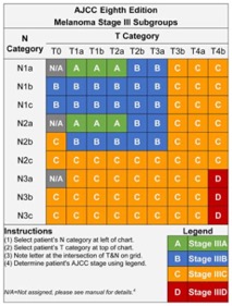
Based on AJCC8 staging criteria, the pathologic tumor stage would be considered pT2b because the thickness is between 1.0-2.0 mm with ulceration. Answers A and C are incorrect because although the thickness is between 1.0mm and 2.0 mm there is ulceration present making pT2b the correct answer. Answer E is incorrect because the thickness is not >2.0 mm- 4.0 mm, making pT3b incorrect. The nodal disease stage would be N2a because the sentinel lymph node biopsy was positive for two microscopic lymph nodes. There were not lymph nodes detected clinically so the nodal disease stage would not be N2b. Based on the tumor stage and the nodal stage the AJCC8 stage would be IIIb as shown in the table below. Answer D is not correct because the Stage is IIIb, not IIIa.
According to NCCN guidelines, sentinel lymph node biopsy is generally not recommended for < 0.8 mm. For tumors with a thickness of 0.8-1.0 mm sentinel lymph node biopsy should be discussed and considered. High risk features to consider for these patients include ulceration, mitoses, and lymphovascular invasion. Sentinel lymph node biopsy should also be discussed and offered to patients with a thickness of > 1.0 mm.
References
- Gershenwald JE, Scolyer RA, Hess KR, et al. Melanoma staging: Evidence‐based changes in the American Joint Committee on Cancer eighth edition cancer staging manual. CA: A Cancer Journal for Clinicians. 2017;67(6):472-492. doi:10.3322/caac.21409
- Coit DG, Thompson JA, Algazi A, et al. Melanoma, Version 2.2016, NCCN Clinical Practice Guidelines in Oncology. Journal of the National Comprehensive Cancer Network : JNCCN. 2016;14(4):450-473.


