
May 2024 Case Study
Authors: Sara Abdel Azim, MS1,2, Kamaria Nelson, MD1, Karl Saardi, MD1
- Department of Dermatology, George Washington University School of Medicine and Health Sciences
- Georgetown University School of Medicine
A 63-year-old female with a past medical history of substance use disorder and breast cancer status post bilateral mastectomy presented with large, violaceous, hemorrhagic purpuric plaques on the chest and bilateral axilla (Fig 1). She had a fever of 104 F and heart rate of 140 bpm. She was admitted to the hospital for atrial fibrillation with rapid ventricular response and was found to be thrombocytopenic with a positive blood culture for streptococcus pyogenes. Coagulation studies revealed a prolonged PTT, an elevated INR level and a low fibrinogen level.
Which of the following is the diagnosis?
A.) Anti-phospholipid syndrome
B.) Calciphylaxis
C.) ANCA associated vasculitis
D.) Purpura fulminans
E.) Warfarin-induced skin necrosis
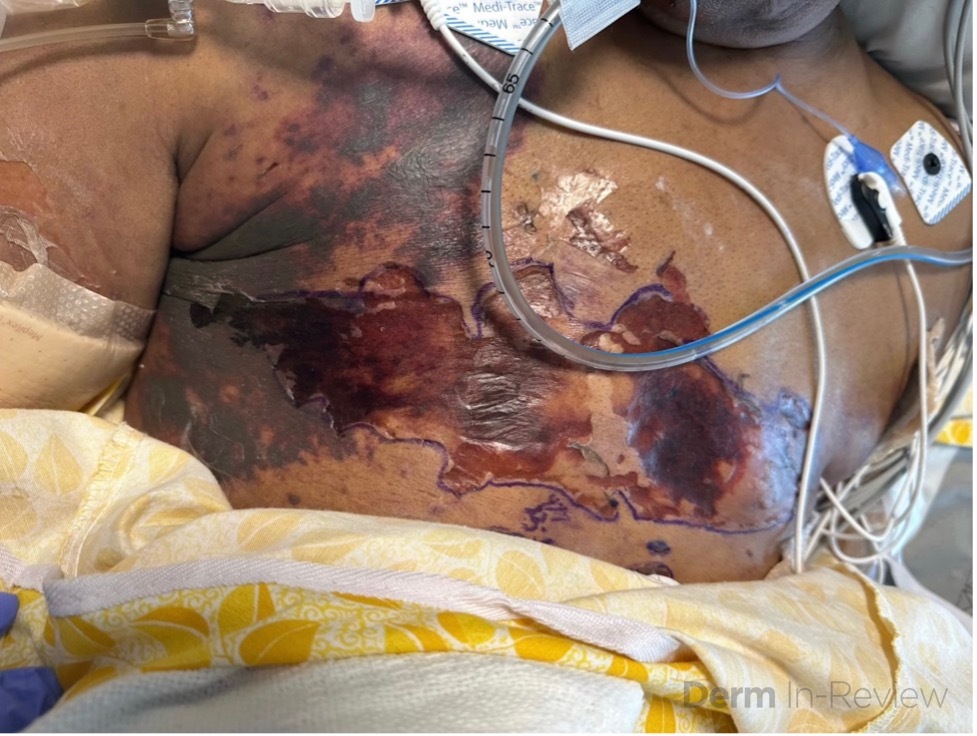
Correct Answer: D – Purpura fulminans
Explanation/Literature review:
Based on the patient’s manifestation of large, hemorrhagic purpuric plaques, acute systemic infection, and concurrent findings of disseminated intravascular coagulation on laboratory tests, she was diagnosed with purpura fulminans (choice D).
Purpura fulminans is a dermatologic emergency involving dysfunction of hemostasis, leading to a hypercoagulable state and ultimately disseminated intravascular coagulation and dermal vascular thrombosis.1 Patients present with ecchymotic skin lesions that may progress to gangrene and result in amputation. Acute infectious purpura fulminans is the most common subtype and occurs in the setting of sepsis, with endotoxin producing gram-negative bacteria triggering an acquired consumption of protein C and S and antithrombin III. 2,3 The most common pathogens are meningococcus and streptococcus pneumoniae, though streptococcus pyogenes has also been implicated.4
Antiphospholipid syndrome (APS) (choice A) is an autoimmune disease characterized by the presence of antiphospholipid antibodies, including lupus anticoagulant, anticardiolipin antibody, and anti-B2-glycoprotein antibody, that damage endothelial cells and disrupt procoagulation defenses, promoting thrombosis.5 Livedo reticularis the most frequently observed lesion, though APS can also cause ulcerations, digital gangrene, subungual splinter hemorrhages, superficial venous thrombosis, and thrombocytopenic purpura. APS occurs secondary to autoimmune disease, most commonly systemic lupus erythematosus (40% of secondary cases)6 or can be primary. Though infections such as borrelia burgdorferi, treponema, HIV, and leptospira have been implicated in the induction of antiphospholipid antibody formation, septicemia is not a common cause of APS and associated disseminated intravascular coagulation is a less common outcome(choice A).7
Calciphylaxis (choice B) is caused by cutaneous arteriolar calcification that leads to subsequent tissue ischemia.8 It presents as violaceous or erythematous, painful cutaneous lesions or subcutaneous nodules and nonhealing wounds, commonly involving adipose-dense regions such as the abdomen, thighs, and buttocks. Calciphylaxis most commonly presents in patients with end stage renal disease but can also occur in patients with acute renal failure, normal renal function, or early stages of chronic kidney disease.
Antineutrophilic cytoplasmic antibody (ANCA) (choice C) associated vasculitis are a heterogenous group of autoimmune conditions that cause inflammation of blood vessels.9 The most common cutaneous manifestation is palpable purpura, though presentation may consist of polymorphic lesions on a background of livedo reticularis.10 This group of conditions has multisystemic effects, often involving the kidneys, sinopulmonary tract, gastrointestinal tract, and lungs, depending on subtype. Infection should be excluded before making the diagnosis.
Warfarin-induced skin necrosis (choice E) occurs in individuals under warfarin treatment with a thrombophilic history or after initiating warfarin therapy with a large loading dose or without concomitant heparin
References
- Bektas F, Soyuncu S. Idiopathic purpura fulminans. Am J Emerg Med. May 2011;29(4):475.e5-6. doi:10.1016/j.ajem.2010.04.022
- Perera T, Murphy-Lavoie H. Purpura Fulminans. StatPearls [Internet]. Treasure Island (FL): StatPearls Publishing; 2023.
- Kim MC, Patel J. Recognition and Management of Acute Purpura Fulminans: A Case Report of a Complication of Neisseria meningitidis Bacteremia. Cureus. Mar 04 2021;13(3):e13704. doi:10.7759/cureus.13704
- Okuzono S, Ishimura M, Kanno S, et al. Streptococcus pyogenes-purpura fulminans as an invasive form of group A streptococcal infection. Ann Clin Microbiol Antimicrob. Jul 09 2018;17(1):31. doi:10.1186/s12941-018-0282-9
- Magdaleno-Tapial J, Valenzuela-Oñate C, Pitarch-Fabregat J, et al. Purpura fulminans-like lesions in antiphospholipid syndrome with endothelial C3 deposition. JAAD Case Rep. Oct 2018;4(9):956-958. doi:10.1016/j.jdcr.2018.09.006
- Bustamant J, Goyal A, Singhal M. Antiphospholipid Syndrome. Treasure Island (FL): StatPearls Publishing. Accessed May 2, 2024. https://www.ncbi.nlm.nih.gov/books/NBK430980/
- Asherson RA, Espinosa G, Cervera R, et al. Disseminated intravascular coagulation in catastrophic antiphospholipid syndrome: clinical and haematological characteristics of 23 patients. Ann Rheum Dis. Jun 2005;64(6):943-6. doi:10.1136/ard.2004.026377
- Westphal S, Plumb T. Calciphylaxis. Treasure Island (FL): StatPearls Publishing. Accessed May 2, 2024. https://www.ncbi.nlm.nih.gov/books/NBK519020/
- Qasim A, Patel J. ANCA Positive Vasculitis. Treasure Island (FL): StatPearls Publishing. Accessed May 2, 2024. https://www.ncbi.nlm.nih.gov/books/NBK554372/