January 2021 Case Study
by Angela Hou, MD
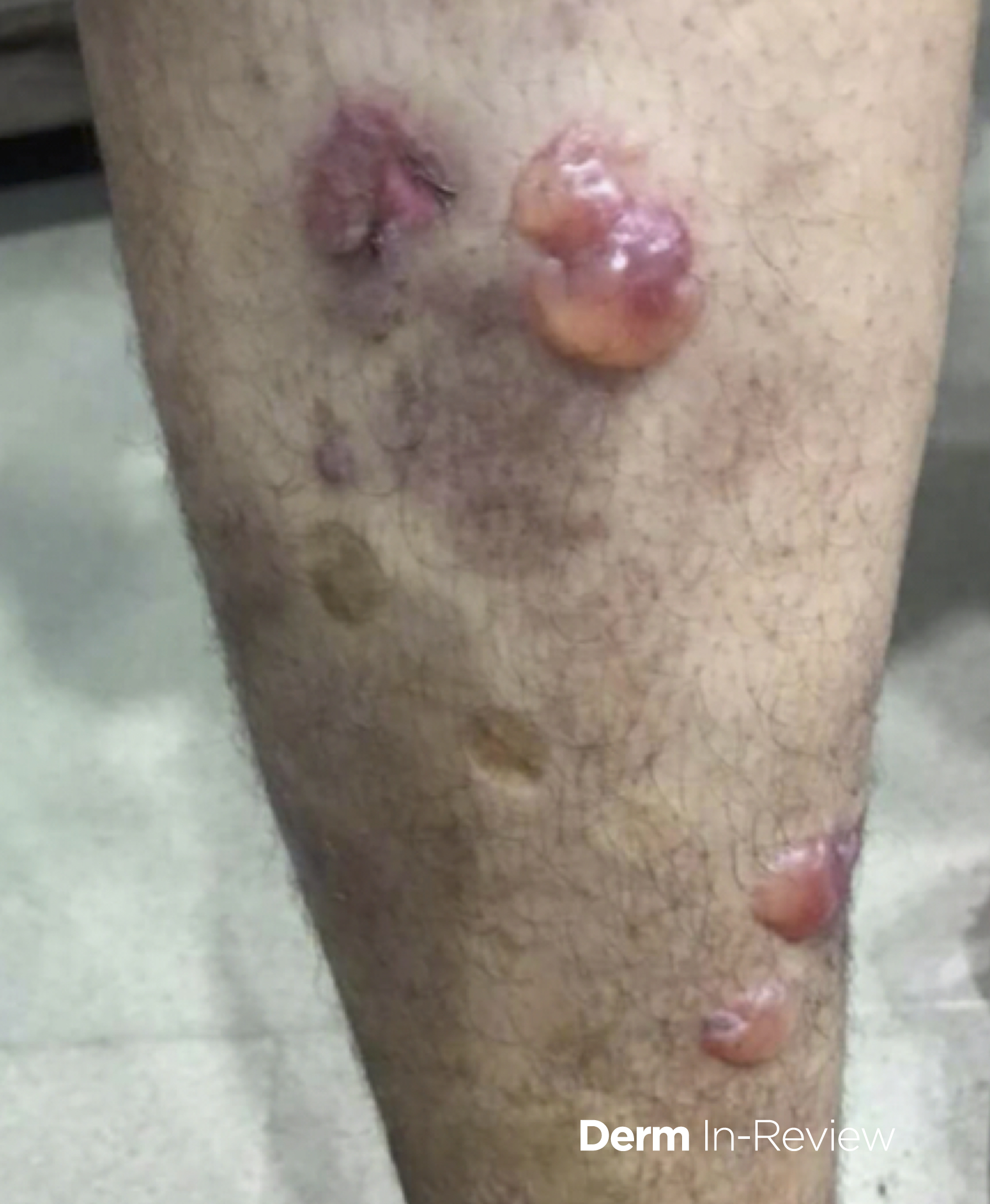
A 51-year-old man presents to clinic with an ongoing rash x1 month. He reports tense blisters on his trunk and lower extremities (Figure 1). He has not used any medications for these lesions. You take a perilesional biopsy for direct immunofluorescence (DIF) and a biopsy for salt split skin immunofluorescence (IMF). DIF shows linear IgG and C3 along the dermal-epidermal junction, and the salt split skin IMF shows IgG on the blister roof.
What is the most likely diagnosis?
A.) Bullous pemphigoid
B.) Linear IgA Disease
C.) Epidermolysis bullosa acquisita
D.) Bullous systemic lupus erythematosus
E.) Pemphigus vulgaris
Correct answer: A) Bullous Permphigoid
This patient presents with a subepidermal blistering disease, which often requires direct immunofluorescence and salt split skin immunofluorescence to differentiate between the various diseases. Multiple subepidermal blistering diseases present with linear IgG and C3 along the dermal-epidermal junction, including bullous pemphigoid, lichen planus pemphigoides, epidermolysis bullosa acquisita, cicatricial pemphigoid, and bullous systemic lupus.
However, in this case, only bullous pemphigoid has both linear IgG and C3 as well as salt split skin IMF that targets the roof of the blister.
Linear IgA disease has linear IgA along the basement membrane instead of IgG and C3. Epidermolysis bullosa acquisita and bullous systemic lupus erythematosus have linear IgG and C3 deposition along the basement membrane but salt split skin IMF has IgG along the floor of the blister. Pemphigus vulgaris is intercellular IgG and C3, not along the basement membrane zone.
References
James, W., Elston, D., Treat, J., Rosenbach, M. and Neuhaus, I., 2020. Andrews’ Diseases of the Skin. 13th ed. Edinburg: Elsevier.