2026 Case Studies – Derm In-Review

June 2026 Case Study

By 2026 Case Studies

June 2026 Case Study

Dillon Nussbaum, MD1

  1. Department of Dermatology, George Washington University School of Medicine and Health Sciences

Patient History   

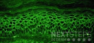
A 58-year-old woman presents with several months of painful oral erosions followed by the development of flaccid bullae on the chest and scalp. Physical examination demonstrates erosions on the buccal mucosa and a positive Nikolsky sign. Direct immunofluorescence demonstrates intercellular IgG and C3 deposition in a reticular “chicken wire” pattern throughout the epidermis (Figure 1). Due to the severity of her disease, she is started on systemic corticosteroids along with a first-line steroid-sparing agent. Which of the following best describes the mechanism by which this steroid-sparing therapy improves her disease?

Figure 1.


  1. Inhibition of IL-17 signaling and neutrophil recruitment
  2. Depletion of CD20-positive B lymphocytes, leading to reduced pathogenic autoantibody production
  3. Blockade of TNF-alpha mediated keratinocyte apoptosis
  4. Inhibition of calcineurin-dependent T-cell activation and IL-2 transcription
  5. Neutralization of circulating IgE antibodies bound to mast cells


Correct Answer: B

Explanation/Literature review:

This patient’s clinical presentation strongly suggests pemphigus vulgaris (PV), an autoimmune blistering disorder characterized by painful mucosal erosions, flaccid bullae, and intercellular “chicken wire” deposition of IgG and C3 on direct immunofluorescence. The pathogenesis of PV is driven by IgG autoantibodies directed primarily against desmoglein (DSG) 3 and, in many patients, DSG1. These desmosomal cadherins are critical for keratinocyte adhesion within the epidermis. Loss of adhesion between keratinocytes results in acantholysis and intraepidermal suprabasal blister formation.

Rituximab has emerged as the preferred first-line steroid-sparing therapy for moderate-to-severe pemphigus vulgaris. Rituximab is a chimeric monoclonal antibody directed against CD20, a surface antigen expressed on mature B lymphocytes. Binding of rituximab to CD20 leads to B-cell depletion through complement-mediated cytotoxicity, antibody-dependent cellular cytotoxicity, and induction of apoptosis. By reducing autoreactive B cells, rituximab decreases the production of pathogenic anti-desmoglein autoantibodies responsible for disease activity.

Multiple studies have demonstrated the efficacy of rituximab in PV. A landmark randomized controlled trial showed that rituximab combined with short-term prednisone achieved significantly higher rates of complete remission off therapy compared with prednisone alone, leading to FDA approval and adoption as first-line therapy. Subsequent systematic reviews and meta-analyses have confirmed high remission rates, reduced cumulative corticosteroid exposure, and improved long-term disease control with rituximab therapy.

The incorrect answer choices represent therapies targeting alternative immune pathways. IL-17 inhibition is used in psoriasis, calcineurin inhibition suppresses T-cell activation, TNF-alpha blockade is used in inflammatory diseases such as hidradenitis suppurativa, and anti-IgE therapy is used in allergic disease. None directly address the pathogenic B-cell–mediated autoantibody production central to pemphigus vulgaris.


References:

  1. Joly, P., Maho-Vaillant, M., Prost-Squarcioni, C., Hebert, V., Houivet, E., Calbo, S., Caillot, F., Golinski, M. L., Labeille, B., Picard-Dahan, C., Paul, C., Richard, M. A., Bouaziz, J. D., Duvert-Lehembre, S., Bernard, P., Caux, F., Alexandre, M., Ingen-Housz-Oro, S., Vabres, P., Delaporte, E., … French study group on autoimmune bullous skin diseases (2017). First-line rituximab combined with short-term prednisone versus prednisone alone for the treatment of pemphigus (Ritux 3): a prospective, multicentre, parallel-group, open-label randomised trial. Lancet (London, England)389(10083), 2031–2040.
  2. Werth, V. P., Joly, P., Mimouni, D., Maverakis, E., Caux, F., Lehane, P., Gearhart, L., Kapre, A., Pordeli, P., Chen, D. M., & PEMPHIX Study Group (2021). Rituximab versus Mycophenolate Mofetil in Patients with Pemphigus Vulgaris. The New England journal of medicine384(24), 2295–2305.
  3. Murrell, D. F., & Sprecher, E. (2017). Rituximab and short-course prednisone as the new gold standard for new-onset pemphigus vulgaris and pemphigus foliaceus. The British journal of dermatology, 177(5), 1143–1144.
  4. Didona, D., Maglie, R., Eming, R., & Hertl, M. (2019). Pemphigus: Current and Future Therapeutic Strategies. Frontiers in immunology, 10, 1418.

April 2026 Case Study

By 2026 Case Studies

April 2026 Case Study

Robin Picavia, MD1

  1. Department of Dermatology, George Washington University School of Medicine and Health Sciences

Patient History

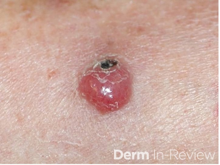
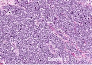
A 72-year-old man with a recent diagnosis of acute myeloid leukemia, colon cancer (in remission) presents with an asymptomatic eruption of multiple violaceous papules and nodules on the trunk and extremities. He denies pain, pruritus, or burning. A punch biopsy demonstrates a dense dermal infiltrate of atypical mononuclear cells. The patient is diagnosed with Leukemia cutis.

Question

Which of the following statements regarding this condition is most accurate?

A. It is most associated with acute lymphoblastic leukemia
B. It typically demonstrates a dense neutrophilic infiltrate on histopathology
C. It may precede the diagnosis of systemic leukemia and is associated with a poorer prognosis
D. It is confined to the epidermis with prominent epidermotropism
E. It is best treated with topical corticosteroids alone

Correct Answer: C. It may precede the diagnosis of systemic leukemia and is associated with a poorer prognosis

Explanation/Literature review:

Leukemia cutis is defined as the infiltration of the skin by malignant leukocytes in patients with underlying leukemia, most commonly acute myeloid leukemia (AML), particularly monocytic or myelomonocytic subtypes (e.g., M4/M5). Clinically, lesions are variable but classically present as firm, asymptomatic papules, nodules, or plaques that are erythematous, violaceous, or skin-colored. They may be localized or widespread and commonly involve the trunk, extremities, and face.

Histopathologically, leukemia cutis demonstrates a dense dermal or subcutaneous infiltrate of atypical mononuclear cells (leukemic blasts), often with sparing of the epidermis (Grenz zone). The cytomorphology depends on the leukemia subtype, and immunohistochemical staining (e.g., myeloperoxidase, CD43, CD68, CD117) is frequently required to confirm myeloid lineage and distinguish it from mimickers such as lymphoma or reactive processes. Correlation with peripheral blood and bone marrow findings is essential for diagnosis.

A key board relevant concept is that leukemia cutis may precede, coincide with, or follow the diagnosis of systemic leukemia. In some patients, cutaneous involvement is the first manifestation of disease (aleukemic leukemia cutis). Importantly, its presence is associated with a worse prognosis, reflecting aggressive disease biology, higher tumor burden, and increased likelihood of extramedullary involvement. Reported survival is significantly reduced compared to patients without skin involvement.

Management is directed at the underlying leukemia, typically with systemic chemotherapy or targeted therapies depending on cytogenetic and molecular features. Cutaneous lesions generally improve with systemic disease control; localized therapies (e.g., radiation) may be considered in select cases but are not first-line.

Answer A is incorrect because leukemia cutis is more strongly associated with AML than acute lymphoblastic leukemia. Answer B describes Sweet syndrome, which shows a dense neutrophilic infiltrate and typically presents with tender plaques and systemic symptoms such as fever. Answer D is incorrect because epidermotropism is characteristic of Cutaneous T-cell lymphoma, not leukemia cutis, which primarily involves the dermis and subcutis. Answer E is incorrect because topical therapies alone are insufficient; treatment must target the systemic malignancy.

References

  1. Gray A, Awethe Z, Himed. Morphologic and Histopathologic Characteristics of Leukemia Cutis. Journal of the American Academy of Dermatology, 91AB260
  2. Robak E, Braun M, Robak T. Leukemia Cutis-The Current View on Pathogenesis, Diagnosis, and Treatment. Cancers (Basel). 2023 Nov 13;15(22):5393. doi: 10.3390/cancers15225393. PMID: 38001655; PMCID: PMC10670312.
  3. Nahm WJ, Juarez M, Abdul-Hay M, Bhatt A, Meehan SA, Shvartsbeyn M. Leukemia Cutis in Relapsed Acute Myeloid Leukemia: A Call for Distinct Classification. Am J Case Rep. 2024 May 18;25:e943577. doi: 10.12659/AJCR.943577. PMID: 38760926; PMCID: PMC11117435.

March 2026 Case Study

By 2026 Case Studies

March 2026 Case Study

Nidhi Shah, MD1 and Emily Murphy, MD1

  1. Department of Dermatology, George Washington University School of Medicine and Health Sciences

Patient History

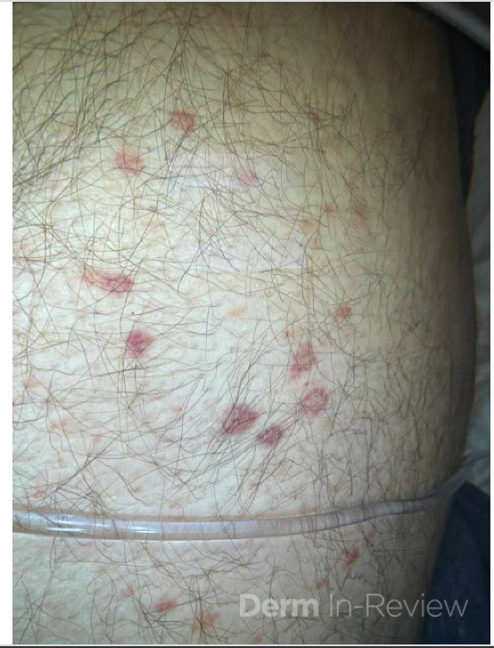
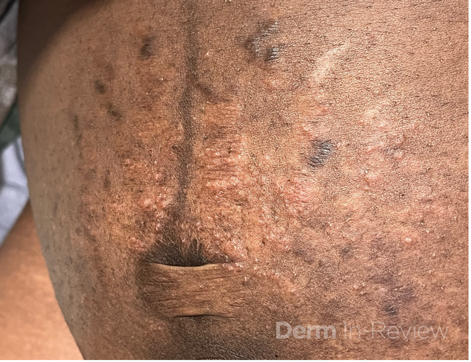
A 28-year-old G1P0 woman at 38 weeks’ gestation presents with a 1-week history of intensely pruritic rash on the abdomen. She reports the rash is limited to the abdomen, denies mucosal involvement, fevers, or prior similar episodes.

On examination, there are erythematous urticarial papules coalescing into plaques within the abdominal striae. The periumbilical region is spared.

Figure 1:


Which of the following fetal risks is most strongly associated with this condition?

  1. Increased risk of stillbirth
    B. Preterm delivery and small-for-gestational-age infant
    C. Neonatal blistering
    D. No increased fetal risk
    E. Neonatal hypothyroidism


Correct Answer: (D) – no increased fetal risk

This patient’s presentation is most consistent with Polymorphic Eruption of Pregnancy (PEP) or Pruritic Urticarial Papules and Plaques of Pregnancy (PUPPP), a benign dermatosis that typically occurs in the third trimester, most often in primiparous women. The eruption characteristically begins within the abdominal striae and spares the umbilicus, presenting as intensely pruritic urticarial papules and plaques. As the name suggests, there can be variable clinical presentations beyond urticaria, including vesicles, diffuse erythema, targetoid lesions, or eczematous plaques. Importantly, polymorphic eruption of pregnancy is not associated with placental dysfunction, autoantibody formation, or systemic maternal disease. As a result, it does not increase the risk of stillbirth, preterm delivery, fetal growth restriction, or neonatal skin disease. PEP usually does not reoccur with future pregnancies. Management is symptomatic (topical steroids and/or antihistamines may be used), and no change in obstetric management is required.

Choice A, increased risk of stillbirth, is most strongly associated with intrahepatic cholestasis of pregnancy. In this condition, patients experience intense pruritus—often involving the palms and soles—without primary inflammatory skin lesions. Elevated serum bile acids are linked to fetal arrhythmias and stillbirth, which is why early delivery is often considered. Management involves reducing serum bile acids using ursodeoxycholic acid. The presence of primary urticarial papules and plaques in this patient makes cholestasis unlikely.

Choice B, preterm delivery and small-for-gestational-age infant, is associated with pemphigoid gestationis, an autoimmune subepidermal blistering disease caused by autoantibodies targeting BP180 (type XVII collagen). Pemphigoid gestationis typically begins in the periumbilical region with urticarial plaques and may progress to vesicles and tense bullae. It carries risks of placental involvement, preterm birth, and fetal growth restriction. Sparing of the periumbilical area and lack of vesicles/bullae argues against this diagnosis.

Choice C, neonatal blistering, can also occur in pemphigoid gestationis due to transplacental transfer of pathogenic IgG autoantibodies. A small percentage of neonates develop transient blisters after birth. Because polymorphic eruption of pregnancy is not antibody-mediated, this complication does not occur.

Choice E, neonatal hypothyroidism, is not associated with any of the common pregnancy-specific dermatoses and serves as a distractor.

References:

  1. Ambros-Rudolph CM. Pregnancy dermatoses. In: Bolognia J, Schaffer JV, Cerroni L, eds. Dermatology. 5th ed. Elsevier; 2025:480-490.


February 2026 Case Study

By 2026 Case Studies

February 2026 Case Study

Caroline Clark, MD1

  1. Department of Dermatology, George Washington University School of Medicine and Health Sciences

Patient History
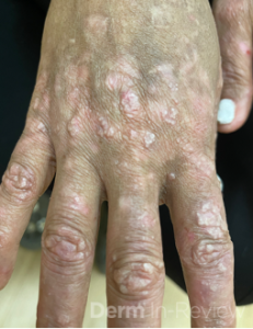
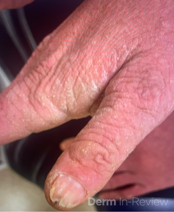
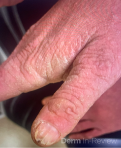
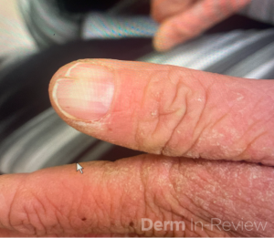
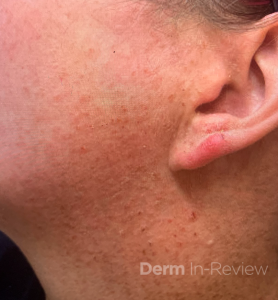
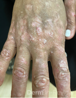
A 19-year-old woman presents to dermatology clinic with a 3-year history of a progressively worsening rash on her neck, chest and hands, and nail changes. The rash is mildly itchy, has a strong odor, and worsens during the summer months when she is sweating or spending time outdoors. She has tried over-the-counter acne treatments and moisturizers without improvement.

Her father reports having similar skin problems that began in his teenage years.

Laboratory studies including complete blood count, comprehensive metabolic panel, and thyroid function tests are within normal limits.

A gene mutation affecting which of the following processes is most likely responsible for this patient’s skin findings?

A) Keratinocyte proliferation and differentiation

B) Degradation of keratin through proteolytic enzymes 

C) Keratinocyte Adhesion

D) Endoplasmic Reticulum calcium transport

E) Golgi Apparatus calcium transport


Correct Answer: D

Explanation/Literature Review 

Darier disease is an autosomal dominant genodermatosis caused by mutations in the ATP2A2 gene, which encodes the sarco/endoplasmic reticulum (ER) calcium ATPase type 2 (SERCA2) pump (Answer choice D).  This genetic defect impairs intracellular calcium homeostasis, leading to desmosome breakdown, acantholysis, and abnormal keratinization.1

The disease typically manifests in the second decade of life with greasy, yellow-brown keratotic papules and plaques in a seborrheic distribution (face, chest, back) and flexural regions, often accompanied by a characteristic malodor.1,2  Nearly all patients develop nail abnormalities including red and white longitudinal streaks with V-shaped notches at the distal edges, and many have palmoplantar pits and oral mucosal papules.

Histologically, the hallmark features are suprabasilar acantholysis with corps ronds (enlarged keratinocytes with fragmented nuclei in the spinous layer) and corps grains (small oval cells in the stratum corneum).2

The disease follows a chronic relapsing course with exacerbations triggered by heat, humidity, UV radiation, mechanical trauma, and infections.1,3

Treatment remains largely symptomatic, with oral retinoids being most effective for extensive disease, though recent evidence suggests targeting the IL-23/IL-17 axis may provide benefit in therapy-resistant cases.1,4

Explanation of Incorrect Answers

Keratinocyte adhesion (Answer choice C) is impaired in Darier disease due to ER stress and abnormal desmosome/adherens junction formation, but this is a downstream consequence rather than the primary molecular defect. Abnormal keratinocyte proliferation and differentiation (Answer choice A) are seen in psoriasis, which affects the nails with pitting, onycholysis, and subungual hyperkeratosis. The nail findings seen in onychomycosis are a result of the dermatophyte’s degradation of keratin through proteolytic enzymes (Answer choice B). Lastly, Hailey-Hailey disease (benign familial pemphigus) is caused by a mutation in ATP2C1 encoding SPCA1, a secretory pathway Ca²⁺-ATPase located in the Golgi apparatus (Answer choice D).  Both Hailey-Hailey and Darier disease are calcium pump disorders with overlapping clinical features including acantholysis, however the keratotic papules and nail V-nicking are more characteristic of Darier disease. 

References

  1. Ettinger M, Kimeswenger S, Deli I, Traxler J, Altrichter S, Noack P, Wikstrom JD, Guenova E, Hoetzenecker W. Darier disease: Current insights and challenges in pathogenesis and management. J Eur Acad Dermatol Venereol. 2025 May;39(5):942-951. doi: 10.1111/jdv.20448. Epub 2024 Nov 28. PMID: 39606894; PMCID: PMC12023721.
  2. Engin B, Kutlubay Z, Erkan E, Tüzün Y. Darier disease: A fold (intertriginous) dermatosis. Clin Dermatol. 2015 Jul-Aug;33(4):448-51. doi: 10.1016/j.clindermatol.2015.04.009. Epub 2015 Apr 9. PMID: 26051059.
  3. Atzmony L, Zagairy F, Mawassi B, et al. Persistent Cutaneous Lesions of Darier Disease and Second-Hit Somatic Variants in ATP2A2 Gene. JAMA Dermatol. 2024;160(5):518–524. doi:10.1001/jamadermatol.2024.0152
  4. Ettinger M, Burner T, Sharma A, Chang YT, Lackner A, Prompsy P, Deli IM, Traxler J, Wahl G, Altrichter S, Langer R, Tsai YC, Varkhande SR, Schoeftner LC, Iselin C, Gratz IK, Kimeswenger S, Guenova E, Hoetzenecker W. Th17-associated cytokines IL-17 and IL-23 in inflamed skin of Darier disease patients as potential therapeutic targets. Nat Commun. 2023 Nov 17;14(1):7470. doi: 10.1038/s41467-023-43210-5. PMID: 37978298; PMCID: PMC10656568.

January 2026 Case Study

By 2026 Case Studies

January 2026 Case Study

Nathaniel Lampley, MD

Patient History 

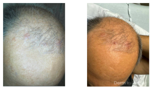
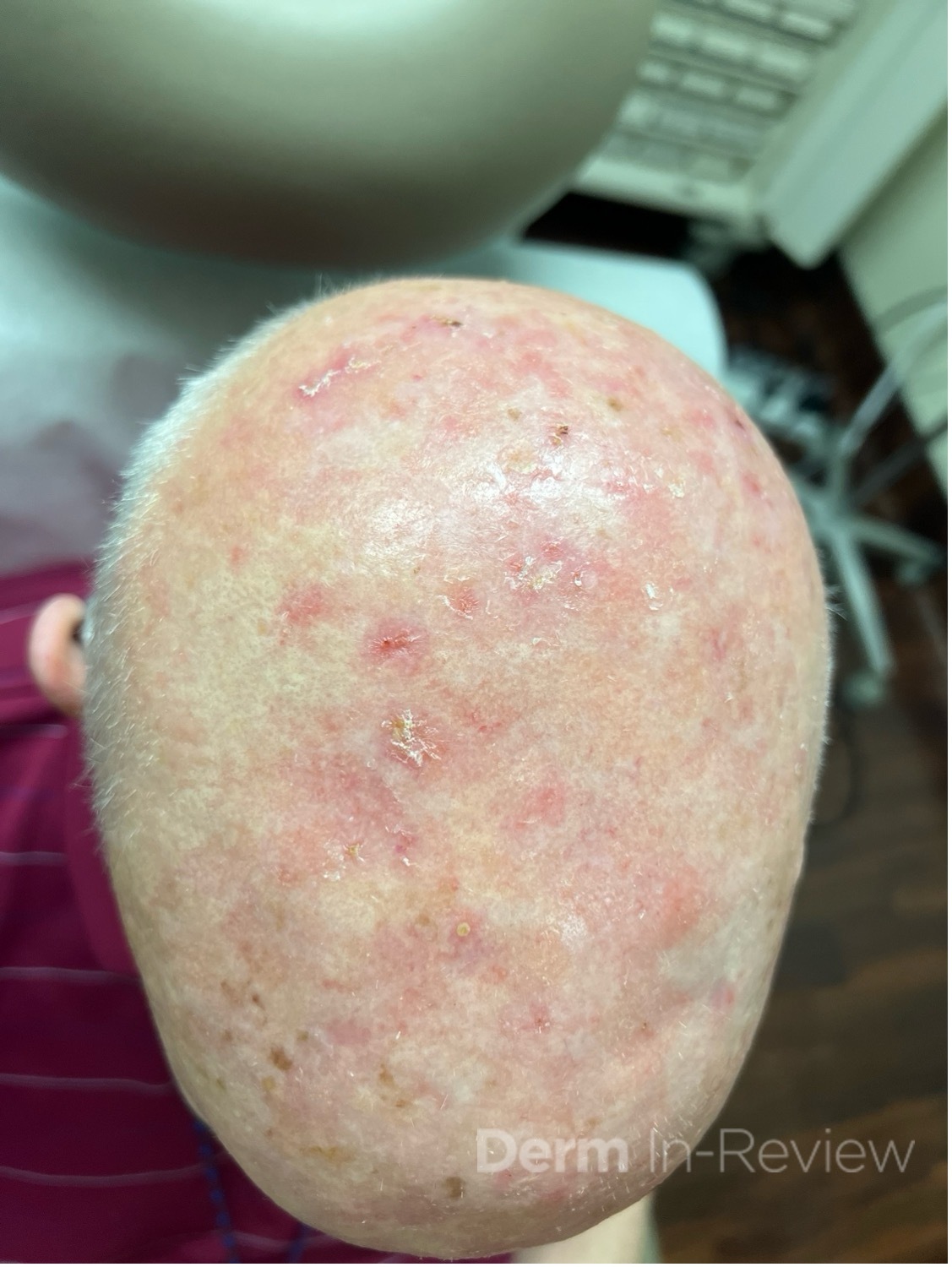
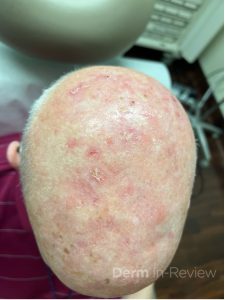
A 60-year-old male presents to your clinic for a skin cancer screening. He has no history of skin cancer or skin cancer treatments, but does admit to a heavy sun exposure history. You identify the pictured scalp lesions during your exam (Figure 1). Given the extent of scalp involvement, you prescribe a topical field therapy. A few days after beginning treatment, he develops rapidly progressive erythema, edema, erosions, and ulceration at the application sites, accompanied by fever, nausea, vomiting, and diarrhea.

Which of the following enzymes is the patient most likely deficient in?

A) Thymidylate synthase

B) Thiopurine methyltransferase

C) Dihydropyrimidine dehydrogenase

D) Inosine monophosphate dehydrogenase


Correct Answer: C

Explanation/Literature Review 

C) This patient presented with actinic keratoses (Figure 1) that were treated with topical 5-fluorouracil (5-FU). A deficiency in dihydropyrimidine dehydrogenase (DPD) can lead to impaired metabolism of 5-fluorouracil (5-FU). DPD is the rate-limiting enzyme responsible for the catabolism of over 80% of administered 5-FU. In patients with partial or complete DPD deficiency, even topical exposure can result in excessive local toxicity due to drug accumulation. 1-3

Reactions typically occur within the first several days of initiating topical therapy and can manifest as severe erosive dermatitis, ulceration, pain that is markedly more intense than the expected inflammatory response seen with standard 5-FU treatment, as well as systemic symptoms.

Recognition of this entity is important, as continued exposure can lead to severe cutaneous injury, and systemic 5-FU administration in patients with undiagnosed DPD deficiency may result in life-threatening toxicity. Management involves immediate cessation of 5-FU, supportive wound care, and avoidance of future fluoropyrimidine exposure. Genetic testing for DPD mutations may be considered.4

Explanation of Incorrect Answers

A) Thymidylate synthase is the intracellular target of 5-fluorouracil, which inhibits DNA synthesis by preventing the conversion of deoxyuridylate to thymidylate. While inhibition of this enzyme mediates the therapeutic effect of 5-FU, deficiency or alteration of thymidylate synthase does not explain excessive toxicity due to impaired drug metabolism.5

B) Thiopurine methyltransferase (TPMT) is involved in the metabolism of thiopurine medications, including azathioprine, 6-mercaptopurine, and 6-thioguanine. Deficiency in TPMT can lead to life-threatening myelosuppression with the administration of these agents, but it has no role in fluoropyrimidine metabolism.6

D) Inosine monophosphate dehydrogenase (IMPDH) is a key enzyme in de novo guanine nucleotide synthesis and is inhibited by medications such as mycophenolate mofetil. Deficiency or inhibition of this enzyme does not affect 5-FU metabolism and is unrelated to fluoropyrimidine toxicity.7

References

  1. Amstutz U, Henricks LM, Offer SM, Barbarino J, Schellens JH, Swen JJ, Klein TE, McLeod HL, Caudle KE, Diasio RB, Schwab M. Clinical Pharmacogenetics Implementation Consortium (CPIC) guideline for dihydropyrimidine dehydrogenase genotype and fluoropyrimidine dosing: 2017 update. Clinical Pharmacology & Therapeutics. 2018 Feb;103(2):210-6.
  2. Ezzeldin H, Diasio R. Dihydropyrimidine dehydrogenase deficiency, a pharmacogenetic syndrome associated with potentially life-threatening toxicity following 5-fluorouracil administration. Clinical colorectal cancer. 2004 Sep 1;4(3):181-9.
  3. Henricks LM, Lunenburg CA, de Man FM, Meulendijks D, Frederix GW, Kienhuis E, Creemers GJ, Baars A, Dezentjé VO, Rosing H, Beijnen JH. DPYD genotype-guided dose individualization of fluoropyrimidine therapy: A prospective safety and cost-analysis on DPYD variants DPYD* 2A, c. 2846A> T, c. 1679T> G and c. 1236G> A. Annals of Oncology. 2018 Oct 1;29:viii150.
  4. Meulendijks D, Henricks LM, Sonke GS, Deenen MJ, Froehlich TK, Amstutz U, Largiadèr CR, Jennings BA, Marinaki AM, Sanderson JD, Kleibl Z. Clinical relevance of DPYD variants c. 1679T> G, c. 1236G> A/HapB3, and c. 1601G> A as predictors of severe fluoropyrimidine-associated toxicity: a systematic review and meta-analysis of individual patient data. The Lancet Oncology. 2015 Dec 1;16(16):1639-50.
  5. Longley DB, Harkin DP, Johnston PG. 5-fluorouracil: mechanisms of action and clinical strategies. Nature reviews cancer. 2003 May 1;3(5):330-8.
  6. Relling MV, Schwab M, WhirlCarrillo M, SuarezKurtz G, Pui CH, Stein CM, Moyer AM, Evans WE, Klein TE, AntillonKlussmann FG, Caudle KE. Clinical pharmacogenetics implementation consortium guideline for thiopurine dosing based on TPMT and NUDT 15 genotypes: 2018 update. Clinical Pharmacology & Therapeutics. 2019 May;105(5):1095-105.
  7. Allison AC, Eugui EM. Mycophenolate mofetil and its mechanisms of action. Immunopharmacology. 2000 May 1;47(2-3):85-118.