July 2026 Case Study
Dillon Nussbaum, MD1
- Department of Dermatology, George Washington University School of Medicine and Health Sciences
Patient History
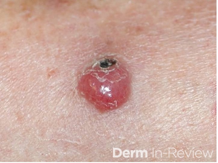
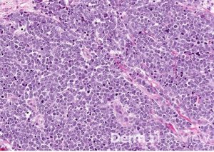
A 71-year-old woman presents with a painless pink eroded papule on the cheek (Figure 1). A shave biopsy is performed and the accompanying pathology is displayed in Figure 2. Based on the histologic findings, which immunohistochemical stain is most helpful to confirm the suspected diagnosis?
Figure 1.
Figure 2.
- CK20
- S100
- TTF-1
- CD3
- CD34
Correct Answer: A. CK20
Explanation/Literature review:
The histologic features strongly suggest Merkel cell carcinoma (MCC), an aggressive primary cutaneous neuroendocrine carcinoma composed of dermal small blue cells with high mitotic activity and characteristic neuroendocrine “salt-and-pepper” chromatin. The absence of an epidermal connection and the clinical presentation of a painless pink papule on sun-exposed skin in an older adult are classic clues. CK20 is the most helpful confirmatory stain because MCC characteristically demonstrates perinuclear dot-like positivity, a staining pattern that is highly characteristic of this tumor in approximately 95% of cases.
TTF-1 can differentiate MCC from metastatic small cell carcinoma of the lung, because small cell carcinoma is typically TTF-1 positive and CK20 negative, whereas MCC is usually CK20 positive and TTF-1 negative. The remaining options are less helpful because S100 is associated with melanoma and other neural crest–derived tumors, CD3 marks T lymphocytes, and CD34 highlights vascular endothelium and certain spindle cell neoplasms such as dermatofibrosarcoma protuberans. None of these markers demonstrates the characteristic staining pattern needed to confirm Merkel cell carcinoma.
References:
- Becker, J. C., Stang, A., DeCaprio, J. A., Cerroni, L., Lebbé, C., Veness, M., & Nghiem, P. (2017). Merkel cell carcinoma. Nature reviews. Disease primers, 3, 17077.
- Chan, J. K., Suster, S., Wenig, B. M., Tsang, W. Y., Chan, J. B., & Lau, A. L. (1997). Cytokeratin 20 immunoreactivity distinguishes Merkel cell (primary cutaneous neuroendocrine) carcinomas and salivary gland small cell carcinomas from small cell carcinomas of various sites. The American journal of surgical pathology, 21(2), 226–234.
- Merkel cell carcinoma. (n.d.). Www.pathologyoutlines.com. https://www.pathologyoutlines.com/topic/skintumornonmelanocyticmerkelcell.html
- Sakata, Y., Inaba, Y., Kunimoto, K., Kaminaka, C., Yamamoto, Y., Iwahashi, Y., Murata, S. I., Asamura, S., & Jinnin, M. (2021). The clinical significance of cytokeratin 20 staining pattern in Merkel cell carcinoma. Drug discoveries & therapeutics, 15(3), 162–165.