Courtney Cruickshank – Derm In-Review

June 2026 Case Study

By 2026 Case Studies

June 2026 Case Study

Dillon Nussbaum, MD1

  1. Department of Dermatology, George Washington University School of Medicine and Health Sciences

Patient History   

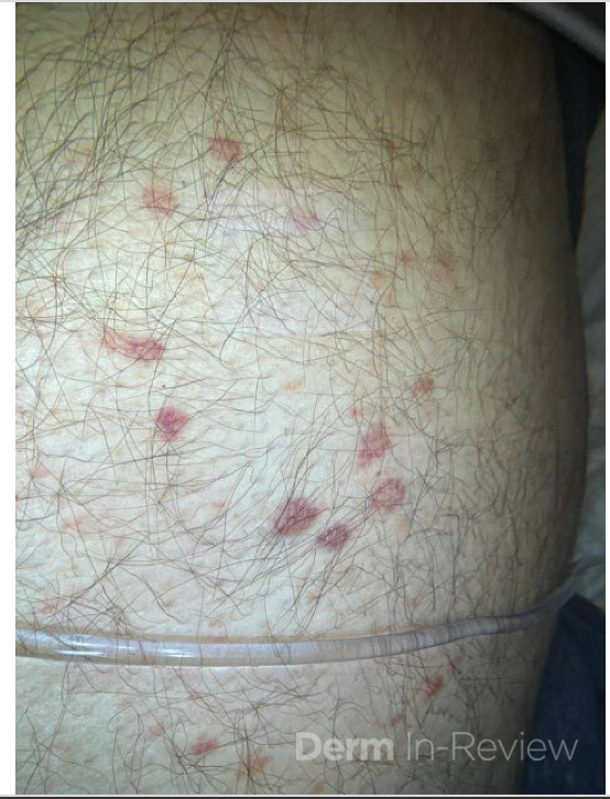
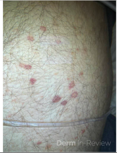
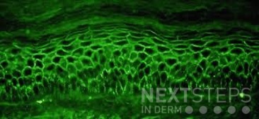
A 58-year-old woman presents with several months of painful oral erosions followed by the development of flaccid bullae on the chest and scalp. Physical examination demonstrates erosions on the buccal mucosa and a positive Nikolsky sign. Direct immunofluorescence demonstrates intercellular IgG and C3 deposition in a reticular “chicken wire” pattern throughout the epidermis (Figure 1). Due to the severity of her disease, she is started on systemic corticosteroids along with a first-line steroid-sparing agent. Which of the following best describes the mechanism by which this steroid-sparing therapy improves her disease?

Figure 1.


  1. Inhibition of IL-17 signaling and neutrophil recruitment
  2. Depletion of CD20-positive B lymphocytes, leading to reduced pathogenic autoantibody production
  3. Blockade of TNF-alpha mediated keratinocyte apoptosis
  4. Inhibition of calcineurin-dependent T-cell activation and IL-2 transcription
  5. Neutralization of circulating IgE antibodies bound to mast cells


Correct Answer: B

Explanation/Literature review:

This patient’s clinical presentation strongly suggests pemphigus vulgaris (PV), an autoimmune blistering disorder characterized by painful mucosal erosions, flaccid bullae, and intercellular “chicken wire” deposition of IgG and C3 on direct immunofluorescence. The pathogenesis of PV is driven by IgG autoantibodies directed primarily against desmoglein (DSG) 3 and, in many patients, DSG1. These desmosomal cadherins are critical for keratinocyte adhesion within the epidermis. Loss of adhesion between keratinocytes results in acantholysis and intraepidermal suprabasal blister formation.

Rituximab has emerged as the preferred first-line steroid-sparing therapy for moderate-to-severe pemphigus vulgaris. Rituximab is a chimeric monoclonal antibody directed against CD20, a surface antigen expressed on mature B lymphocytes. Binding of rituximab to CD20 leads to B-cell depletion through complement-mediated cytotoxicity, antibody-dependent cellular cytotoxicity, and induction of apoptosis. By reducing autoreactive B cells, rituximab decreases the production of pathogenic anti-desmoglein autoantibodies responsible for disease activity.

Multiple studies have demonstrated the efficacy of rituximab in PV. A landmark randomized controlled trial showed that rituximab combined with short-term prednisone achieved significantly higher rates of complete remission off therapy compared with prednisone alone, leading to FDA approval and adoption as first-line therapy. Subsequent systematic reviews and meta-analyses have confirmed high remission rates, reduced cumulative corticosteroid exposure, and improved long-term disease control with rituximab therapy.

The incorrect answer choices represent therapies targeting alternative immune pathways. IL-17 inhibition is used in psoriasis, calcineurin inhibition suppresses T-cell activation, TNF-alpha blockade is used in inflammatory diseases such as hidradenitis suppurativa, and anti-IgE therapy is used in allergic disease. None directly address the pathogenic B-cell–mediated autoantibody production central to pemphigus vulgaris.


References:

  1. Joly, P., Maho-Vaillant, M., Prost-Squarcioni, C., Hebert, V., Houivet, E., Calbo, S., Caillot, F., Golinski, M. L., Labeille, B., Picard-Dahan, C., Paul, C., Richard, M. A., Bouaziz, J. D., Duvert-Lehembre, S., Bernard, P., Caux, F., Alexandre, M., Ingen-Housz-Oro, S., Vabres, P., Delaporte, E., … French study group on autoimmune bullous skin diseases (2017). First-line rituximab combined with short-term prednisone versus prednisone alone for the treatment of pemphigus (Ritux 3): a prospective, multicentre, parallel-group, open-label randomised trial. Lancet (London, England)389(10083), 2031–2040.
  2. Werth, V. P., Joly, P., Mimouni, D., Maverakis, E., Caux, F., Lehane, P., Gearhart, L., Kapre, A., Pordeli, P., Chen, D. M., & PEMPHIX Study Group (2021). Rituximab versus Mycophenolate Mofetil in Patients with Pemphigus Vulgaris. The New England journal of medicine384(24), 2295–2305.
  3. Murrell, D. F., & Sprecher, E. (2017). Rituximab and short-course prednisone as the new gold standard for new-onset pemphigus vulgaris and pemphigus foliaceus. The British journal of dermatology, 177(5), 1143–1144.
  4. Didona, D., Maglie, R., Eming, R., & Hertl, M. (2019). Pemphigus: Current and Future Therapeutic Strategies. Frontiers in immunology, 10, 1418.

Krazy Kodachromes: Twenty Ninth Installment

By Krazy Kodachrome

Twenty Ninth Installment

Date: May 30th, 2024

Guests:

Dr. Emily Murphy
Dr. Kamaria Nelson
Dr. Colleen Cotton
Dr. Adam Friedman

Krazy Kodachromes: Thirtieth Installment

By Krazy Kodachrome

Thirtieth Installment

Date: August 24, 2024
Guests:
Dr. Yasmine Kirkorian
Dr. Austin Cox
Dr. Adam Friedman

Krazy Kodachromes: Thirty Third Installment

By Krazy Kodachrome

Thirty Third Installment

Date: May 29, 2025

Guests:
Dr. Adam Friedman
Dr. Aubrey Montebello
Dr. Erik Kumetz
Dr. Alexis Carrington
Dr. Adam Rosenfeld

April 2026 Case Study

By 2026 Case Studies

April 2026 Case Study

Robin Picavia, MD1

  1. Department of Dermatology, George Washington University School of Medicine and Health Sciences

Patient History

A 72-year-old man with a recent diagnosis of acute myeloid leukemia, colon cancer (in remission) presents with an asymptomatic eruption of multiple violaceous papules and nodules on the trunk and extremities. He denies pain, pruritus, or burning. A punch biopsy demonstrates a dense dermal infiltrate of atypical mononuclear cells. The patient is diagnosed with Leukemia cutis.

Question

Which of the following statements regarding this condition is most accurate?

A. It is most associated with acute lymphoblastic leukemia
B. It typically demonstrates a dense neutrophilic infiltrate on histopathology
C. It may precede the diagnosis of systemic leukemia and is associated with a poorer prognosis
D. It is confined to the epidermis with prominent epidermotropism
E. It is best treated with topical corticosteroids alone

Correct Answer: C. It may precede the diagnosis of systemic leukemia and is associated with a poorer prognosis

Explanation/Literature review:

Leukemia cutis is defined as the infiltration of the skin by malignant leukocytes in patients with underlying leukemia, most commonly acute myeloid leukemia (AML), particularly monocytic or myelomonocytic subtypes (e.g., M4/M5). Clinically, lesions are variable but classically present as firm, asymptomatic papules, nodules, or plaques that are erythematous, violaceous, or skin-colored. They may be localized or widespread and commonly involve the trunk, extremities, and face.

Histopathologically, leukemia cutis demonstrates a dense dermal or subcutaneous infiltrate of atypical mononuclear cells (leukemic blasts), often with sparing of the epidermis (Grenz zone). The cytomorphology depends on the leukemia subtype, and immunohistochemical staining (e.g., myeloperoxidase, CD43, CD68, CD117) is frequently required to confirm myeloid lineage and distinguish it from mimickers such as lymphoma or reactive processes. Correlation with peripheral blood and bone marrow findings is essential for diagnosis.

A key board relevant concept is that leukemia cutis may precede, coincide with, or follow the diagnosis of systemic leukemia. In some patients, cutaneous involvement is the first manifestation of disease (aleukemic leukemia cutis). Importantly, its presence is associated with a worse prognosis, reflecting aggressive disease biology, higher tumor burden, and increased likelihood of extramedullary involvement. Reported survival is significantly reduced compared to patients without skin involvement.

Management is directed at the underlying leukemia, typically with systemic chemotherapy or targeted therapies depending on cytogenetic and molecular features. Cutaneous lesions generally improve with systemic disease control; localized therapies (e.g., radiation) may be considered in select cases but are not first-line.

Answer A is incorrect because leukemia cutis is more strongly associated with AML than acute lymphoblastic leukemia. Answer B describes Sweet syndrome, which shows a dense neutrophilic infiltrate and typically presents with tender plaques and systemic symptoms such as fever. Answer D is incorrect because epidermotropism is characteristic of Cutaneous T-cell lymphoma, not leukemia cutis, which primarily involves the dermis and subcutis. Answer E is incorrect because topical therapies alone are insufficient; treatment must target the systemic malignancy.

References

  1. Gray A, Awethe Z, Himed. Morphologic and Histopathologic Characteristics of Leukemia Cutis. Journal of the American Academy of Dermatology, 91AB260
  2. Robak E, Braun M, Robak T. Leukemia Cutis-The Current View on Pathogenesis, Diagnosis, and Treatment. Cancers (Basel). 2023 Nov 13;15(22):5393. doi: 10.3390/cancers15225393. PMID: 38001655; PMCID: PMC10670312.
  3. Nahm WJ, Juarez M, Abdul-Hay M, Bhatt A, Meehan SA, Shvartsbeyn M. Leukemia Cutis in Relapsed Acute Myeloid Leukemia: A Call for Distinct Classification. Am J Case Rep. 2024 May 18;25:e943577. doi: 10.12659/AJCR.943577. PMID: 38760926; PMCID: PMC11117435.