September 2026 Case Study
Dillon Nussbaum, MD 1
1. Department of Dermatology, George Washington University School of Medicine and Health
Sciences
Patient History
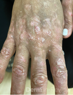
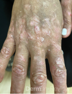
A 57-year-old woman presents with the findings seen in Figure 1, which are bilaterally on the dorsal hands and forearms. She has mild arthralgias and a history of intermittent oral ulcers. A past biopsy report, which only provides direct immunofluorescence, shows deposition of multiple antibodies along the basement membrane zone that localize predominantly to the dermal side on salt-split skin. Autoantibodies against which of the following structures is most directly responsible for this patient’s findings?
Figure 1.
A. Desmoglein 1
B. BP180
C. Type VII collagen
D. Laminin-332
E. Desmoplakin
Correct Answer: C. Type VII collagen
The combination of a neutrophil-rich subepidermal blister, multiple antibodies at the dermoepidermal junction, dermal-side staining on salt-split skin, and systemic features of systemic lupus erythematosus strongly suggests a diagnosis of bullous lupus erythematosus. The key structural target of bullous lupus erythematosus is type VII collagen, which forms the anchoring fibrils that extend from the lamina densa into the papillary dermis. Autoantibodies against type VII collagen impair this attachment, producing separation beneath the lamina densa and ultimately subepidermal vesicles, which tend to be tense.
The incorrect answers represent other cellular structures involved in the dermoepidermal adhesion apparatus. Desmoglein 1 (A) and desmoplakin (E) are desmosomal proteins involvedi n keratinocyte-to-keratinocyte adhesion and are associated with intraepidermal blistering. BP180 (B) is a hemidesmosomal transmembrane protein targeted in bullous pemphigoid. Laminin-332 (D) is a basement membrane component involved in epithelial adhesion but does not form the anchoring fibrils themselves. The dermal-side salt-split pattern is the major clue directing you toward an antigen below the lamina densa, particularly type VII collagen.
References:
1. Grabell, D. A., Matthews, L. A., Yancey, K. B., & Chong, B. F. (2015). Detection of Type VII
Collagen Autoantibodies Before the Onset of Bullous Systemic Lupus
Erythematosus. JAMA dermatology, 151(5), 539–543.
https://doi.org/10.1001/jamadermatol.2014.4409
2. Sebaratnam, D. F., & Murrell, D. F. (2011). Bullous systemic lupus
erythematosus. Dermatologic clinics, 29(4), 649–653.
https://doi.org/10.1016/j.det.2011.06.002