
Case Studies are a resource of Derm In-Review,
brought to you by our educational partner
Featured Case Study

Question
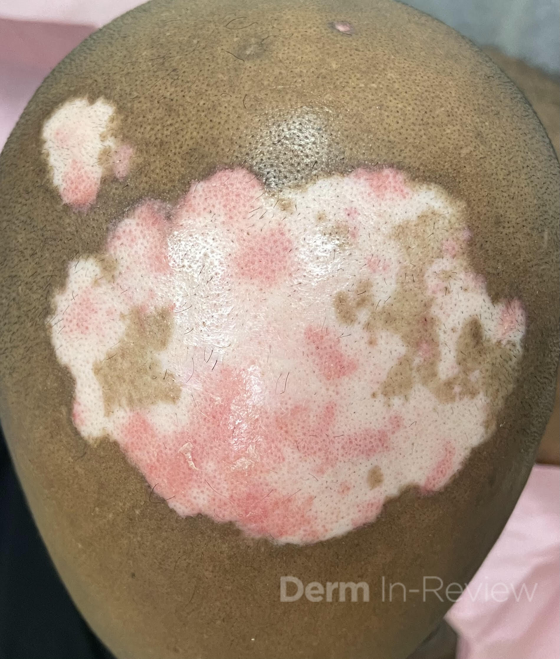
A 42-year-old man presents with several well-demarcated, erythematous plaques with central atrophic hypopigmentation, peripheral hyperpigmentation, and adherent scale on his scalp, back, and cheeks for several months (Figure 1). He notes mild pruritus and hair loss in the affected areas but denies systemic symptoms. A punch biopsy is obtained and is significant for interface dermatitis and mucin deposition.
Which of the following ophthalmologic findings may develop in a small percentage of patients taking the first-line systemic therapy for this condition longitudinally?
1. Kayser-Fleischer rings
2. Cataracts
3. Central scotoma
4. Retinal detachment
5. Optic neuritis
Correct Answer
Correct Answer: C
Explanation
Histopathologic findings of interface dermatitis and mucin deposition along with clinical findings of atrophic hypopigmented plaques with peripheral hyperpigmentation are consistent with a diagnosis of discoid lupus erythematosus (DLE).1 The first-line systemic agent for widespread or refractory DLE is hydroxychloroquine, which inhibits Toll-like receptors 7 and 9 and downregulates the interferon mediated inflammatory cascade implicated in DLE. Although generally well-tolerated, hydroxychloroquine carries a risk of retinal toxicity, particularly with long-term use or dosing above 5 mg/kg/day. Hydroxychloroquine accumulates in the retinal pigment epithelium and can cause a central scotoma, which presents as a focal blurry or blind spot at the center of the visual field, and is typically irreversible.
Studies indicate the incidence of central scotoma in patients taking hydroxychloroquine ranges from 0.5% to 7.5% after 5 years of therapy, with most cases being mild, and the variability depending on diagnostic criteria. Baseline and annual ophthalmologic exams, including visual field testing and optical coherence tomography, are recommended after 5 years of therapy or earlier in higher-risk patients.2-4
DLE commonly occurs in sun exposed areas with a predilection for the conchal bowls, often resolves with scarring and alopecia, and is worsened by tobacco use. Topical therapies include corticosteroids and non-steroidal anti-inflammatory agents in addition to daily photoprotection. Hydroxychloroquine is first line for both systemic lupus erythematosus (SLE) and DLE and other systemic immunosuppressants are frequently used in conjunction with hydroxychloroquine when appropriate. Janus kinase inhibitors are increasingly being utilized off label for both DLE and SLE. While uncommon, DLE can be associated with SLE, and if so, monoclonal antibodies such as belimumab or anifrolumab can be utilized.5,6 Incorrect answers include Kayser-Fleischer rings, which are dark copper colored rings on the periphery of the iris seen in Wilson’s disease. Cataracts, or clouding of the lens, is associated with corticosteroid use among other etiologies, but has not been associated with the use of hydroxychloroquine. Retinal detachment and optic neuritis are unrelated to hydroxychloroquine use.
References
1. McDaniel B, Sukumaran S, Koritala T, et al. Discoid Lupus Erythematosus. [Updated 2023 Aug 28]. In: StatPearls [Internet]. Treasure Island (FL): StatPearls Publishing; 2025 Jan-.
2. Ahn S. J. (2024). Classification of Hydroxychloroquine Retinopathy: A Literature Review and Proposal for Revision. Diagnostics (Basel, Switzerland), 14(16), 1803.
3. Marshall, E., Robertson, M., Kam, S., Penwarden, A., Riga, P., & Davies, N. (2021). Prevalence of hydroxychloroquine retinopathy using 2018 Royal College of Ophthalmologists diagnostic criteria. Eye (London, England), 35(1), 343–348.
4. Melles, R. B., Jorge, A. M., Marmor, M. F., Zhou, B., Conell, C., Niu, J., McCormick, N., Zhang, Y., & Choi, H. K. (2023). Hydroxychloroquine Dose and Risk for Incident Retinopathy : A Cohort Study. Annals of internal medicine, 176(2), 166–173.
5. Abduelmula, A., Sood, S., Mufti, A., Hinek, A., & Yeung, J. (2023). Management of cutaneous lupus erythematosus with Janus kinase inhibitor therapy: An evidence-based review. Journal of the American Academy of Dermatology, 89(1), 130–131.
6. Merola, J. F., Prystowsky, S. D., Iversen, C., Gomez-Puerta, J. A., Norton, T., Tsao, P., Massarotti, E., Schur, P., Bermas, B., & Costenbader, K. H. (2013). Association of discoid lupus erythematosus with other clinical manifestations among patients with systemic lupus erythematosus. Journal of the American Academy of Dermatology, 69(1), 19–24.
1
1