
Case Studies are a resource of Derm In-Review,
brought to you by our educational partner
Featured Case Study

Question
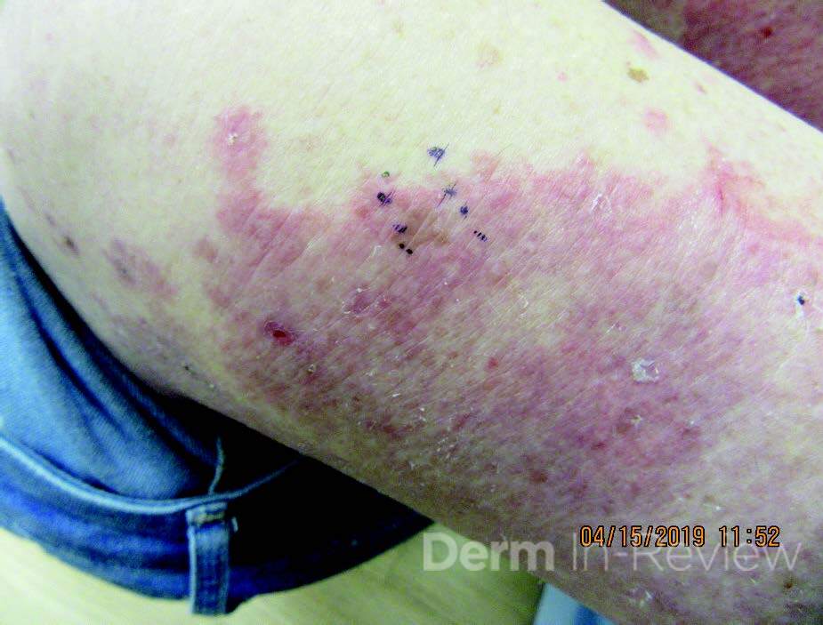
Patient is an 84-year-old woman who presents with new pruritic urticarial plaques on her legs, thighs, and upper back. They have been expanding for 2 weeks and are recalcitrant to topical steroids. A biopsy was performed, which showed spongiosis and abundant eosinophils in the dermis. DIF shows linear deposition of C3 and IgG along the dermal-epidermal junction.
Which of the following conditions have the same antigenic target as in the disease seen in this patient?
1. Herpes gestationis
2. Epidermolysis bullosa acquisita
3. Bullous lupus erythematosus
4. Ocular-predominant mucous membrane pemphigoid
5. IgA pemphigus
Correct Answer
Correct Answer: A. Herpes gestationis
Explanation
The patient in this question stem has bullous pemphigoid. Patients with bullous pemphigoid often present with initially urticarial or eczematous plaques, then develop tense bullae. Biopsy shows subepidermal bulla with eosinophilic infiltrate, and the characteristic DIF nding includes linear IgG and C3 along the basement membrane. The antigenic targets for bullous pemphigoid are BP180 (BPAg2) and BP230 (BPAg1).
Herpes gestationis, also known as pemphigoid gestationis, has autoantibodies against BP180 like bullous pemphigoid. This is a rare vesiculobullous eruption that occurs in patients in the second or third trimester of pregnancy. Like bullous pemphigoid it initially starts as extremely pruritic urticarial papules and plaques that progress into tense blisters. The lesions are commonly distributed around the umbilicus. It is important to educate patients that this commonly recurs during future pregnancies, and that the severity often increases with subsequent pregnancies.
Epidermolysis bullosa acquisita is an acquired autoimmune blistering disease with antibodies targeting collagen type VII. Bullous lupus erythematosus also has antibodies directed at collagen type VII, but tends to improve rapidly to treatment with dapsone, whereas epidermolysis bullosa acquisita is usually recalcitrant to treatment. While mucous membrane pemphigoid can have autoantibodies targeting BP180, in the ocular-predominant subtype targets 4 integrin specically. IgA pemphigus presents in middle-aged to elderly patients with flaccid vesicles and pustules in an annular or circinate pattern. It has two subtypes: subcorneal pustular dermatosis, in which the target antigen is desmocollin 1, and intraepidermal neutrophilic type, with target antigens desmoglein 1 and 3.
References
References
1. Culton DA, Liu Z, Diaz LA. Bullous pemphigoid. In: Kang S, Amagai M, Bruckner AL, Enk AH, Margolis DJ, McMichael AJ,
Orringer JS, eds. Fitzpatrick’s Dermatology. 9th ed. New York, NY: McGraw-Hill Education; 2019.
2. Lipozen i J, Ljubojevic S, Bukvi -Mokos Z. Pemphigoid gestationis. Clin Dermatol. 2012 Jan-Feb;30(1):51-5.
3. Ambros-Rudolph CM. Chapter 27. Pregnancy dermatoses. In: Bolognia JL, Schaffer JV, Cerroni L, eds. Dermatology. 4th ed.
Philadelphia, PA: Elsevier; 2017:472-483.
4. Chi CC, Wang SH, Charles-Holmes R, et al. Pemphigoid gestationis: early onset and blister formation are associated with
adverse pregnancy outcomes. Br J Dermatol. 2009 Jun;160(6):1222-8
1
1