Date: May 2026
Guests:
Dr. Mary Maiberger
Dr. Leonardo Tjahjono
Dr. Nidhi Shah
Dr. Nagasai Adusumilli
Thirty Sixth Installment
July 2026 Case Study
Dillon Nussbaum, MD1
- Department of Dermatology, George Washington University School of Medicine and Health Sciences
Patient History
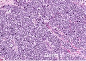
A 71-year-old woman presents with a painless pink eroded papule on the cheek (Figure 1). A shave biopsy is performed and the accompanying pathology is displayed in Figure 2. Based on the histologic findings, which immunohistochemical stain is most helpful to confirm the suspected diagnosis?
Figure 1.
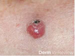
Figure 2.
- CK20
- S100
- TTF-1
- CD3
- CD34
Correct Answer: A. CK20
Explanation/Literature review:
The histologic features strongly suggest Merkel cell carcinoma (MCC), an aggressive primary cutaneous neuroendocrine carcinoma composed of dermal small blue cells with high mitotic activity and characteristic neuroendocrine “salt-and-pepper” chromatin. The absence of an epidermal connection and the clinical presentation of a painless pink papule on sun-exposed skin in an older adult are classic clues. CK20 is the most helpful confirmatory stain because MCC characteristically demonstrates perinuclear dot-like positivity, a staining pattern that is highly characteristic of this tumor in approximately 95% of cases.
TTF-1 can differentiate MCC from metastatic small cell carcinoma of the lung, because small cell carcinoma is typically TTF-1 positive and CK20 negative, whereas MCC is usually CK20 positive and TTF-1 negative. The remaining options are less helpful because S100 is associated with melanoma and other neural crest–derived tumors, CD3 marks T lymphocytes, and CD34 highlights vascular endothelium and certain spindle cell neoplasms such as dermatofibrosarcoma protuberans. None of these markers demonstrates the characteristic staining pattern needed to confirm Merkel cell carcinoma.
References:
- Becker, J. C., Stang, A., DeCaprio, J. A., Cerroni, L., Lebbé, C., Veness, M., & Nghiem, P. (2017). Merkel cell carcinoma. Nature reviews. Disease primers, 3, 17077.
- Chan, J. K., Suster, S., Wenig, B. M., Tsang, W. Y., Chan, J. B., & Lau, A. L. (1997). Cytokeratin 20 immunoreactivity distinguishes Merkel cell (primary cutaneous neuroendocrine) carcinomas and salivary gland small cell carcinomas from small cell carcinomas of various sites. The American journal of surgical pathology, 21(2), 226–234.
- Merkel cell carcinoma. (n.d.). Www.pathologyoutlines.com. https://www.pathologyoutlines.com/topic/skintumornonmelanocyticmerkelcell.html
- Sakata, Y., Inaba, Y., Kunimoto, K., Kaminaka, C., Yamamoto, Y., Iwahashi, Y., Murata, S. I., Asamura, S., & Jinnin, M. (2021). The clinical significance of cytokeratin 20 staining pattern in Merkel cell carcinoma. Drug discoveries & therapeutics, 15(3), 162–165.
June 2026 Case Study
Dillon Nussbaum, MD1
- Department of Dermatology, George Washington University School of Medicine and Health Sciences
Patient History
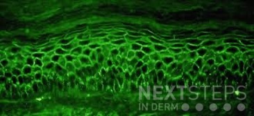
A 58-year-old woman presents with several months of painful oral erosions followed by the development of flaccid bullae on the chest and scalp. Physical examination demonstrates erosions on the buccal mucosa and a positive Nikolsky sign. Direct immunofluorescence demonstrates intercellular IgG and C3 deposition in a reticular “chicken wire” pattern throughout the epidermis (Figure 1). Due to the severity of her disease, she is started on systemic corticosteroids along with a first-line steroid-sparing agent. Which of the following best describes the mechanism by which this steroid-sparing therapy improves her disease?
Figure 1.
- Inhibition of IL-17 signaling and neutrophil recruitment
- Depletion of CD20-positive B lymphocytes, leading to reduced pathogenic autoantibody production
- Blockade of TNF-alpha mediated keratinocyte apoptosis
- Inhibition of calcineurin-dependent T-cell activation and IL-2 transcription
- Neutralization of circulating IgE antibodies bound to mast cells
Correct Answer: B
Explanation/Literature review:
This patient’s clinical presentation strongly suggests pemphigus vulgaris (PV), an autoimmune blistering disorder characterized by painful mucosal erosions, flaccid bullae, and intercellular “chicken wire” deposition of IgG and C3 on direct immunofluorescence. The pathogenesis of PV is driven by IgG autoantibodies directed primarily against desmoglein (DSG) 3 and, in many patients, DSG1. These desmosomal cadherins are critical for keratinocyte adhesion within the epidermis. Loss of adhesion between keratinocytes results in acantholysis and intraepidermal suprabasal blister formation.
Rituximab has emerged as the preferred first-line steroid-sparing therapy for moderate-to-severe pemphigus vulgaris. Rituximab is a chimeric monoclonal antibody directed against CD20, a surface antigen expressed on mature B lymphocytes. Binding of rituximab to CD20 leads to B-cell depletion through complement-mediated cytotoxicity, antibody-dependent cellular cytotoxicity, and induction of apoptosis. By reducing autoreactive B cells, rituximab decreases the production of pathogenic anti-desmoglein autoantibodies responsible for disease activity.
Multiple studies have demonstrated the efficacy of rituximab in PV. A landmark randomized controlled trial showed that rituximab combined with short-term prednisone achieved significantly higher rates of complete remission off therapy compared with prednisone alone, leading to FDA approval and adoption as first-line therapy. Subsequent systematic reviews and meta-analyses have confirmed high remission rates, reduced cumulative corticosteroid exposure, and improved long-term disease control with rituximab therapy.
The incorrect answer choices represent therapies targeting alternative immune pathways. IL-17 inhibition is used in psoriasis, calcineurin inhibition suppresses T-cell activation, TNF-alpha blockade is used in inflammatory diseases such as hidradenitis suppurativa, and anti-IgE therapy is used in allergic disease. None directly address the pathogenic B-cell–mediated autoantibody production central to pemphigus vulgaris.
References:
- Joly, P., Maho-Vaillant, M., Prost-Squarcioni, C., Hebert, V., Houivet, E., Calbo, S., Caillot, F., Golinski, M. L., Labeille, B., Picard-Dahan, C., Paul, C., Richard, M. A., Bouaziz, J. D., Duvert-Lehembre, S., Bernard, P., Caux, F., Alexandre, M., Ingen-Housz-Oro, S., Vabres, P., Delaporte, E., … French study group on autoimmune bullous skin diseases (2017). First-line rituximab combined with short-term prednisone versus prednisone alone for the treatment of pemphigus (Ritux 3): a prospective, multicentre, parallel-group, open-label randomised trial. Lancet (London, England), 389(10083), 2031–2040.
- Werth, V. P., Joly, P., Mimouni, D., Maverakis, E., Caux, F., Lehane, P., Gearhart, L., Kapre, A., Pordeli, P., Chen, D. M., & PEMPHIX Study Group (2021). Rituximab versus Mycophenolate Mofetil in Patients with Pemphigus Vulgaris. The New England journal of medicine, 384(24), 2295–2305.
- Murrell, D. F., & Sprecher, E. (2017). Rituximab and short-course prednisone as the new gold standard for new-onset pemphigus vulgaris and pemphigus foliaceus. The British journal of dermatology, 177(5), 1143–1144.
- Didona, D., Maglie, R., Eming, R., & Hertl, M. (2019). Pemphigus: Current and Future Therapeutic Strategies. Frontiers in immunology, 10, 1418.
Twenty Seventh Installment
Date: August 2023
Guests:
Dr. Adam Friedman
Dr. Pooja Sodha
Dr. Cyrelle Finan
This Krazy Kodachrome is sponsored by Pfizer.
Twenty Eighth Installment
Date: November 2023
Guests:
Dr. James Cox
Dr. Nicholas Logemann
Dr. Luke Bloomquist
Twenty Ninth Installment
Date: May 30th, 2024
Guests:
Dr. Emily Murphy
Dr. Kamaria Nelson
Dr. Colleen Cotton
Dr. Adam Friedman
Thirtieth Installment
Date: August 24, 2024
Guests:
Dr. Yasmine Kirkorian
Dr. Austin Cox
Dr. Adam Friedman
Thirty First Installment
Date: October 31st, 2024
Guests:
Dr. Adam Friedman
Dr. Austin Cox
Dr. Karl Saardi
Thirty Second Installment
Date: January 30, 2025
Guests:
Dr. Emily Murphy
Dr. Emily Murphy
Dr. Adam Friedman
Dr. Karl Saardi
Thirty Third Installment
Date: May 29, 2025
Guests:
Dr. Adam Friedman
Dr. Aubrey Montebello
Dr. Erik Kumetz
Dr. Alexis Carrington
Dr. Adam Rosenfeld